🦵 Anatomy · BIO 252
Your BIO 252 course, all fourteen modules, written out as study material instead of a file list. Each one opens with the one idea that makes the rest make sense, then the structures, then a comparison table and the traps. Open only the module you are revising.
Module 1 ⚗️ Intro to A&P & chemistry What anatomy is, and the chemistry the rest of the course sits on
💡 The big idea
Anatomy is structure. Physiology is function. This course is the structure one — what a thing is and where it sits, not what it does moment to moment.
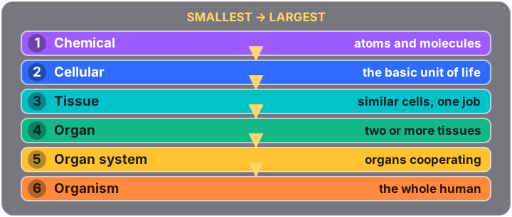
Everything in the body is built in levels, and each level is made of the one below it. That single sentence is the spine of Module 1.
🔍 Two ways to look
| Kind | What it studies | Example |
|---|---|---|
| Gross (macroscopic) | Structures you can see with the naked eye | The chambers of the heart |
| Microscopic | Structures needing a microscope — cells (cytology) and tissues (histology) | Cardiac muscle cells on a slide |
| Developmental | How the body changes across a lifetime, including embryology | How the heart tube becomes four chambers |
🧱 Levels of organization, smallest first
⚛️ Subatomic particles
| Particle | Charge | Where |
|---|---|---|
| Proton | +1 | Nucleus |
| Neutron | 0 | Nucleus |
| Electron | −1 | Orbiting the nucleus |
An ion is an atom whose electrons and protons no longer match, so it carries a charge. Cation = positive (Na+). Anion = negative (Cl−).
🔗 Chemical bonds — transfer, share, or attract
| Bond | What happens to the electrons | Example |
|---|---|---|
| Ionic | Transferred from one atom to another | NaCl |
| Polar covalent | Shared unequally | H2O |
| Non-polar covalent | Shared equally | O2 |
| Hydrogen | Weak attraction between polar molecules | Water to water; holding DNA together |
🧬 The four organic compounds
- Carbohydrates — glucose, fructose, starch
- Lipids — triglycerides, phospholipids, cholesterol
- Proteins — enzymes, hemoglobin, keratin
- Nucleic acids — DNA and RNA
⚠️ Exam trap
Hydrogen bonds are weak individually — but there are so many of them that they are what holds the two strands of DNA together and what gives water its properties. Weak bond does not mean unimportant.
Take it further🧬 The physiology side of this →
Module 2 🧂 The cellular level The membrane that decides, and the organelles that do the work
💡 The big idea
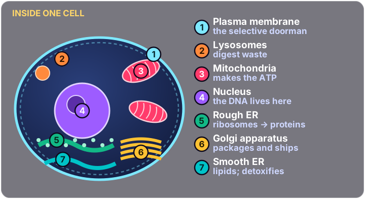
A cell is a factory with a doorman. The membrane decides what comes in and goes out; everything inside is a production line with one job each.
Selectively permeable is the phrase that matters — not “a barrier”, but a barrier that chooses.
🧩 The plasma membrane
Structure: a phospholipid bilayer with proteins embedded in it.
It does five things:
- Physical barrier between inside and outside
- Controls what enters and exits — selectively permeable
- Senses the outside world through receptors
- Anchors the cell to its neighbours
- Gives the cell its shape
🏭 The organelles, one job each
| Organelle | Job | Remember it as |
|---|---|---|
| Nucleus | Holds the DNA; runs the cell | The control centre |
| Mitochondria | Makes ATP | The powerhouse |
| Rough ER | Studded with ribosomes; builds proteins | Rough = Ribosomes |
| Smooth ER | Builds lipids; detoxifies | Smooth has no ribosomes |
| Golgi apparatus | Modifies, packages and ships proteins | The post office |
| Lysosomes | Digestive enzymes; break down waste | The recycling bin |
| Ribosomes | Protein synthesis — on the rough ER or free-floating | The protein factory |
| Cytoskeleton | Structural framework; helps movement | The scaffolding |
⚫ Inside the nucleus
- Nuclear envelope — a double membrane with pores
- Nucleolus — makes ribosomes
- Chromatin — DNA unwound, the working form
- Chromosome — the same DNA condensed, for cell division
🧬 Nucleotides — three parts, always
A nucleotide is the building block of a nucleic acid, and it has exactly three pieces:
| DNA | RNA | |
|---|---|---|
| Sugar | Deoxyribose | Ribose |
| Bases | A, T, C, G | A, U, C, G |
⚠️ Exam trap
Rough ER and smooth ER are the same organelle with and without ribosomes — and that one difference decides the product. Rough builds proteins, smooth builds lipids.
Take it further🧬 The physiology side of this →
Module 3 🧵 Tissues, membranes & skin Name the tissue by its layers and its shape
💡 The big idea
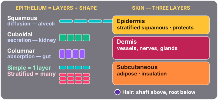
Every epithelial name is two words, and both words are instructions. The first says how many layers, the second says what shape the top cells are.
Simple = one layer, made for passing things through. Stratified = many layers, made for taking a beating.
🪜 Epithelial tissues
| Type | Looks like | Job | Where |
|---|---|---|---|
| Simple squamous | One layer, thin and flat | Diffusion | Alveoli, capillaries |
| Simple cuboidal | One layer, cube-shaped | Secretion, absorption | Kidney tubules |
| Simple columnar | One layer, tall | Absorption | Intestines |
| Stratified squamous | Many layers, flat on top | Protection | Skin, esophagus, vagina |
| Pseudostratified columnar | Looks layered but is not | Secretion, cilia sweep | Trachea |
🧸 Connective tissues
| Tissue | Where | Structure & job |
|---|---|---|
| Areolar | Under the skin | Loose fibres; cushions organs |
| Adipose | Under skin, around organs | Fat cells; stores energy, insulates |
| Reticular | Lymph nodes, spleen | Fibre network; supports immune cells |
| Dense regular | Tendons, ligaments | Parallel collagen — strong in one direction |
| Dense irregular | Dermis | Criss-crossed collagen — strong in many directions |
| Cartilage | Joints, ear, nose | Firm but flexible |
| Bone | Skeleton | Rigid matrix; support and protection |
| Blood | Vessels | A fluid matrix — yes, blood is connective tissue |
🥥 Body membranes
| Membrane | Lines | Secretes |
|---|---|---|
| Mucous | Cavities open to the outside — gut, airway | Mucus |
| Serous | Cavities closed to the outside | Thin serous fluid; reduces friction |
| Cutaneous | The skin itself | — |
| Synovial | Joints | Synovial fluid |
Visceral covers the organ. Parietal lines the cavity wall.
🦵 Skin — three layers, outside in
Hair: the shaft is the visible part above the skin, the root is below it, the follicle surrounds the root, and the bulb is the swollen base where growth happens.
⚠️ Exam trap
Pseudostratified columnar looks like several layers because the nuclei sit at different heights — but every cell touches the basement membrane, so it is a single layer. That is the whole reason it has its own name.
Take it further🖼️ Skin & Wound infographics🎯 Quiz me on skin & burns📖 Psoriasis 🩹
Module 4 🦴 Bone tissue & the skeletal system A build crew, a demolition crew, and three kinds of muscle
💡 The big idea
Bone is living tissue that is constantly being torn down and rebuilt. It is not scaffolding that was poured once. Two cell types are in a permanent argument, and the winner decides whether bone gets denser or thinner.
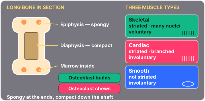
Osteoblast builds. Osteoclast chews.
🦵 Bone cells
| Cell | Job | Where it lives |
|---|---|---|
| Osteoblast | Builds bone | Periosteum, endosteum |
| Osteoclast | Breaks down bone | Bone surfaces |
| Osteocyte | Maintains bone; a retired osteoblast | Trapped in lacunae |
| Osteogenic | Stem cell for the others | Periosteum, marrow |
🪧 Bone shapes
| Shape | Description | Example |
|---|---|---|
| Long | Longer than wide; movement | Femur, humerus |
| Short | Cube-shaped; stability | Carpals, tarsals |
| Flat | Thin and flat; protection | Skull, ribs, sternum |
| Irregular | Complex shapes | Vertebrae |
| Sesamoid | Embedded in a tendon; reduces friction | Patella |
🧿 Compact against spongy
| Compact | Spongy | |
|---|---|---|
| Texture | Dense, solid | Porous, open lattice |
| Where | Outer shell; the diaphysis (shaft) | Inner; the epiphysis (ends) |
| Doing what | Taking the load | Housing marrow, absorbing shock |
💪 The three muscle types
| Type | Structure | Job | Voluntary? |
|---|---|---|---|
| Skeletal | Striated, multinucleated | Moves the skeleton | Yes |
| Cardiac | Striated, branched, one nucleus | Pumps blood | No |
| Smooth | Not striated, spindle-shaped | Moves substances through hollow organs | No |
⚠️ Exam trap
Cardiac muscle is striated, so “striated” is not a safe clue for voluntary. If a stem says striated and branched, it is cardiac every time.
Take it further🖼️ Musculoskeletal infographics🎯 Quiz me on musculoskeletal
Module 5 ⚡ Muscle & nervous tissue How a nerve signal becomes a movement
💡 The big idea
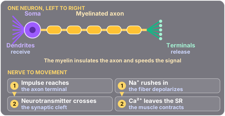
A nerve never touches the muscle it moves. There is always a gap, and the signal has to cross it chemically. That gap is the neuromuscular junction, and almost every drug and disease in this area works by interfering with it.
Electrical → chemical → electrical again.
🧵 Nerve to movement, in order
Calcium is the trigger. No calcium, no cross-bridge, no contraction — which is why calcium turns up in every muscle question eventually.
🧠 The parts of a neuron
| Part | Job |
|---|---|
| Dendrites | Receive signals |
| Soma (cell body) | Holds the nucleus; the metabolic centre |
| Axon | Sends signals away |
| Myelin sheath | Insulates the axon and speeds the signal up |
🌐 Neuron types by shape
| Type | Shape | Where |
|---|---|---|
| Multipolar | Many dendrites, one axon | Most of the CNS; motor neurons |
| Bipolar | One dendrite, one axon | Retina, olfactory epithelium |
| Unipolar | One process that splits | Sensory neurons |
| Pseudounipolar | One process, splitting later | Sensory neurons of the PNS |
| Anaxonic | No distinguishable axon | Brain; function not fully known |
🔗 The synapse, part by part
- Presynaptic neuron — sends
- Axon terminals — where the message is released
- Synaptic vesicles — the sacs holding neurotransmitter
- Synaptic cleft — the gap itself
- Postsynaptic neuron — receives
⚠️ Exam trap
Sodium starts the electrical change; calcium starts the mechanical one. Na+ depolarizes, Ca2+ contracts. Swapping them is the commonest mistake in this module.
Take it further🖼️ Neuro infographics🎯 Quiz me on neurological
Module 6 🧠 Nerves & the nervous system CNS, PNS, the meninges — and all twelve cranial nerves
💡 The big idea
The CNS decides. The PNS carries. Brain and spinal cord are the central nervous system; everything else — cranial nerves, spinal nerves, ganglia — is peripheral.
The cranial nerves are the part this course tests hardest, and the part nursing keeps testing after it.
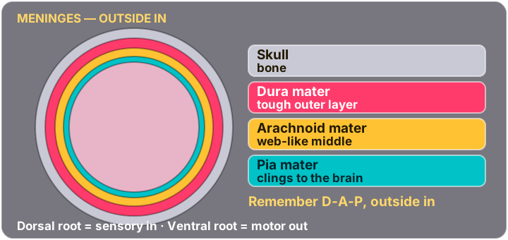
🧿 Meninges, outside in
↔️ Roots: which way does it go
| Root | Carries |
|---|---|
| Dorsal root | Sensory — coming in |
| Ventral root | Motor — going out |
| Spinal nerve | Mixed — both, once they join |
🌌 The cerebellum
Coordinates voluntary movement and maintains posture and balance. It does not start movement — it makes movement smooth. Damage shows up as clumsiness and unsteady gait, not paralysis.
🌟 The twelve cranial nerves
| # | Name | What it does | S / M / B |
|---|---|---|---|
| I | Olfactory | Smell | S |
| II | Optic | Vision | S |
| III | Oculomotor | Most eye movement; pupil constriction; eyelid lift | M |
| IV | Trochlear | Eye down-and-in (superior oblique) | M |
| V | Trigeminal | Facial sensation; chewing; corneal reflex | B |
| VI | Abducens | Eye abduction (lateral rectus) | M |
| VII | Facial | Facial expression; taste front ⅔; tears and saliva | B |
| VIII | Vestibulocochlear | Hearing and balance | S |
| IX | Glossopharyngeal | Taste back ⅓; gag reflex; swallowing | B |
| X | Vagus | Say “aah” — palate and uvula rise; voice; heart, lungs, gut | B |
| XI | Accessory | Shrug shoulders; turn head | M |
| XII | Hypoglossal | Tongue movement | M |
🚨 The two that get confused, and how the exam separates them
IX and X both run to the throat, so a question has to pick one by the test you perform:
- CN X (vagus) — have the client open the mouth and say “aah”. The palate and uvula should rise symmetrically. Also listen to the voice for hoarseness.
- CN IX (glossopharyngeal) — touch the back of the throat with a tongue blade to provoke the gag reflex.
Saying “aah” is vagus. The gag reflex is glossopharyngeal.
🧠 The two mnemonics worth having
Names, I → XII:
On Old Olympus' Towering Tops A Finn And
German Viewed A Hop.
Sensory, motor or both:
Some Say Marry Money But My Brother Says
Big Brains Matter More.
Read the second one against the table above — letter by letter, I through XII.
👀 Sensory receptors
| Receptor | Detects |
|---|---|
| Thermoreceptors | Temperature |
| Nociceptors | Pain |
| Mechanoreceptors | Touch, pressure, vibration |
| Photoreceptors | Light |
| Chemoreceptors | Chemicals — taste and smell |
⚠️ Exam trap
Three different nerves move the eye — III, IV and VI — and the stem tells you which by naming a direction. IV takes the eye down-and-in; VI ab-ducts it outward; III does everything else, plus the pupil.
Take it further🖼️ Neuro infographics🎯 Quiz me on neurological
Module 7 👁️ Special senses The path light takes, and the path sound takes
💡 The big idea
Both special senses are relay races. Nothing detects light or sound directly — each structure hands off to the next, and a question is usually asking you to name one runner.
Learn both pathways in order and most of the module answers itself.
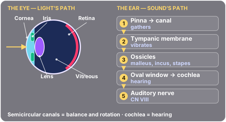
👁️ The eye, front to back
| Structure | Where | Job |
|---|---|---|
| Cornea | Front of the eye | Focuses light as it enters |
| Iris | Around the pupil | Controls how much light gets in |
| Lens | Behind the iris | Fine-focuses light onto the retina |
| Vitreous body | Inside the eye | Holds the eye's shape |
| Retina | Back of the eye | Holds the photoreceptors — where light becomes signal |
| Sclera | Outer coat | Protection and structure — the white of the eye |
The lacrimal gland makes tears, which lubricate and protect the surface.
👂 Sound's path, in order
🌌 Hearing and balance structures
| Structure | Where | Job |
|---|---|---|
| Tympanic membrane | End of the ear canal | Vibrates with sound |
| Ossicles | Middle ear | Transmit vibration — malleus, incus, stapes |
| Eustachian tube | Middle ear to throat | Equalises pressure |
| Cochlea | Inner ear | Hearing |
| Semicircular canals | Inner ear | Rotational balance |
| Ampulla | In the semicircular canals | Detects head rotation |
| Saccule | Vestibule | Balance in the vertical plane |
🧠 Two hooks that carry the module
Ossicles in order: MIS — Malleus (hammer), Incus (anvil), Stapes (stirrup). Outer to inner, and the stapes is the smallest bone in the body.
Cochlea hears, canals balance. Both sit in the inner ear and both report through CN VIII — which is exactly why one nerve is called vestibulo-cochlear.
⚠️ Exam trap
The eustachian tube is the reason a child with a cold gets an ear infection: it is shorter, wider and more horizontal in children, so anything in the throat travels up it easily. That single anatomical fact comes back in peds all year.
Take it further🖼️ Neuro infographics🎯 Quiz me on eyes, ears & sensory
Module 8 🧥 The endocrine system Broadcasting instead of calling, and the receptor that decides who hears
💡 The big idea
A neurotransmitter is a phone call. A hormone is a radio broadcast. One crosses a gap of nanometres to a single target; the other goes into the blood and reaches everything.
Only cells with the right receptor respond — that is what makes a broadcast specific.
💬 Five ways cells talk
| Type | How far |
|---|---|
| Direct | Cell to cell, physically touching |
| Synaptic | Across a synapse, neuron to target |
| Autocrine | The cell signals itself |
| Paracrine | Nearby cells only |
| Endocrine | Through the bloodstream to anywhere |
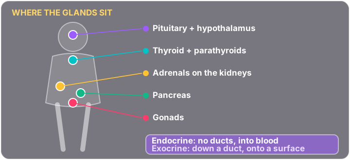
💧 Endocrine against exocrine
| Endocrine | Exocrine | |
|---|---|---|
| Ducts | None | Yes |
| Releases into | Blood | A surface or cavity |
| Examples | Thyroid, adrenals, pituitary | Sweat, saliva, digestive enzymes |
📡 Receptor types
| Receptor | Where | What it does |
|---|---|---|
| Ion channel-linked | Cell membrane | Opens a channel for ions |
| G protein-coupled | Cell membrane | Activates second messengers |
| Enzyme-linked | Cell membrane | Triggers enzyme activity |
| Intracellular | Inside the cell | Binds lipid-soluble hormones — steroids |
A steroid can cross the membrane because it is fat-soluble, so its receptor is waiting inside. Water-soluble hormones cannot get in, so their receptors sit on the surface.
🧠 The posterior pituitary stores two hormones
Oxytocin and ADH (antidiuretic hormone).
The posterior pituitary does not make them — the hypothalamus does, and the posterior pituitary only stores and releases them. That distinction is a favourite question.
⚠️ Exam trap
“Hormones travel in the blood” is true but not the discriminator. If a question asks why only some cells respond, the answer is always the receptor, not the hormone.
Take it further🖼️ Endocrine infographics🎯 Quiz me on endocrine & metabolic
Module 9 🩸 Cardiovascular I — blood & vessels What blood is made of, and which way each vessel runs
💡 The big idea
Arteries and veins are defined by direction, not by oxygen. An artery carries blood away from the heart whatever is in it.
A = Artery = Away. Get that and the pulmonary exception stops being confusing.
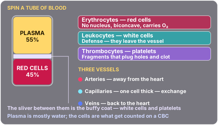
🧂 What is actually in blood
| Fraction | Roughly | Contains |
|---|---|---|
| Plasma | ~55% | Water, proteins, nutrients, hormones, wastes |
| Formed elements | ~45% | Red cells, white cells, platelets |
| Formed element | Job |
|---|---|
| Erythrocytes (RBCs) | Carry oxygen |
| Leukocytes (WBCs) | Immune defence |
| Thrombocytes (platelets) | Clotting |
By count, red cells make up nearly all of the 45%. White cells and platelets together are well under 1%.
↔️ Arteries against veins
| Arteries | Veins | |
|---|---|---|
| Direction | Away from the heart | Toward the heart |
| Walls | Thick and muscular | Thinner |
| Pressure | High | Low |
| Valves | No | Yes — to stop backflow against gravity |
🚨 The pulmonary exception
Usually arteries carry oxygenated blood and veins carry deoxygenated. The pulmonary vessels reverse it:
- Pulmonary arteries carry deoxygenated blood to the lungs
- Pulmonary veins carry oxygenated blood back to the heart
Which is why the definition is direction, never oxygen content.
💓 Where you feel a pulse
| Site | Where |
|---|---|
| Carotid | Neck |
| Brachial | Inner elbow |
| Radial | Wrist — thumb side |
| Femoral | Groin |
A pulse is the artery wall expanding as each heartbeat pushes blood through it. You feel it over arteries, never veins.
⚠️ Exam trap
Blood is a connective tissue — cells suspended in a fluid matrix. It appears in the tissue module and again here, and students who learned it only as “a fluid” miss it both times.
Take it further🎯 Quiz me on hematology & oncology
Module 10 ❤️ Cardiovascular II — the heart Two pumps in one organ, and the wiring that fires them
💡 The big idea
The heart is two pumps side by side. The right side sends blood to the lungs; the left side sends it to the body. That is why the left ventricle has the thickest wall — it pushes against the whole body instead of one organ.
Right goes to the lungs. Left goes to the world.
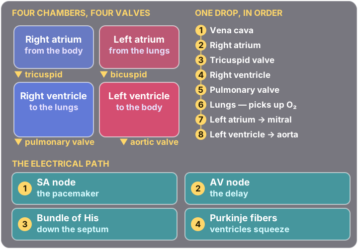
🧭 The four chambers
| Chamber | Receives from | Sends to |
|---|---|---|
| Right atrium | The body, via the vena cava | Right ventricle |
| Right ventricle | Right atrium | The lungs |
| Left atrium | The lungs | Left ventricle |
| Left ventricle | Left atrium | The whole body, via the aorta |
🚪 The four valves
| Valve | Sits between |
|---|---|
| Tricuspid | Right atrium and right ventricle |
| Pulmonary | Right ventricle and pulmonary artery |
| Bicuspid (mitral) | Left atrium and left ventricle |
| Aortic | Left ventricle and aorta |
🔄 The whole circuit, in order
⚡ The conduction system
The AV delay is not a flaw — without it the ventricles would contract while the atria were still filling them.
⚠️ Exam trap
The great vessels are named for where they are going, not what they carry. Pulmonary arteries leave the heart carrying deoxygenated blood. Reading a pathway question with “artery = oxygen” in your head gets it wrong every time.
Take it further🖼️ Cardio infographics🎯 Quiz me on cardiovascular
Module 11 🛡️ Lymphatic & immune systems Fast and generic, or slow and specific — and where each cell is trained
💡 The big idea
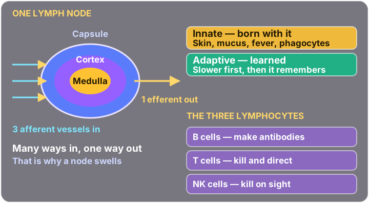
You have two immune systems and they trade speed for precision. Innate is there from birth, responds in minutes, and treats every threat the same. Adaptive takes days the first time, targets one specific thing — and remembers it.
Memory is what makes vaccination possible, and only adaptive immunity has it.
🎓 Where each lymphocyte is trained
| Cell | Matures in |
|---|---|
| B cells | Bone marrow |
| T cells | Thymus |
| NK cells | Bone marrow |
⚖️ Innate against adaptive
| Innate | Adaptive | |
|---|---|---|
| Speed | Immediate | Days, the first time |
| Target | Anything foreign | One specific antigen |
| Memory | None | Yes |
| Players | Skin, mucous membranes, phagocytes, NK cells, inflammation | B cells, T cells, antibodies, memory cells |
Skin and mucous membranes count as innate immunity — they are the first barrier, and an intact one is the cheapest defence the body has.
🧠 Inside a lymph node
| Part | What it is |
|---|---|
| Capsule | The outer wrapping |
| Cortex | Outer region; contains the follicles |
| Medulla | Inner region |
| Afferent vessels | Bring lymph in |
| Efferent vessels | Carry lymph out |
⚠️ Exam trap
A lymphocyte looks unremarkable under a microscope: a large nucleus and very little cytoplasm. If a slide question describes a white cell that is nearly all nucleus, that is a lymphocyte.
Take it further🎯 Quiz me on immune & infection
Module 12 🧤 The respiratory system The tube that conducts, and the sacs that exchange
💡 The big idea
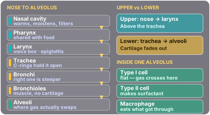
The airway does two different jobs, and only one of them is gas exchange. Everything from the nose down to the bronchioles is plumbing — warming, filtering, conducting. Exchange happens only at the alveoli.
Conducting zone moves air. Respiratory zone trades gas.
🔼 Upper and lower tract
| Tract | Structures |
|---|---|
| Upper | Nose, nasal cavity, pharynx, larynx |
| Lower | Trachea, bronchi, bronchioles, alveoli |
The dividing line is the larynx — above it upper, below it lower.
🌳 Going down the bronchial tree, three things change
| As you go deeper | Direction | Why it matters |
|---|---|---|
| Cartilage | Decreases → disappears | Nothing left to hold the tube open |
| Smooth muscle | Increases | The tube can now clamp shut |
| Diameter | Decreases | Less room to lose |
🦠 The three alveolar cells
| Cell | Job |
|---|---|
| Type I | Gas exchange — thin and flat, most of the surface |
| Type II | Secretes surfactant |
| Alveolar macrophages | Clear debris and pathogens |
Surfactant lowers surface tension so the alveoli do not collapse on themselves at the end of each breath. A premature baby short of it has to fight for every breath — that is respiratory distress syndrome.
⚠️ Exam trap
Type I cells cover most of the alveolar surface but Type II cells are more numerous. “Most of the surface” and “most of the cells” are different questions with different answers.
Take it further🖼️ Respiratory infographics🎯 Quiz me on respiratory
Module 13 🥪 The digestive system One tube from end to end, with helpers plumbed into it
💡 The big idea
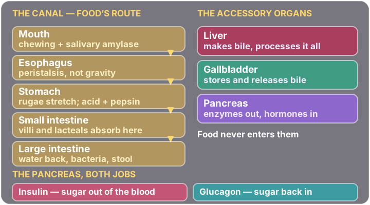
The digestive tract is a single tube, and its whole design problem is surface area. Folds inside folds inside folds — because absorption happens per square centimetre, and a smooth tube would not have nearly enough.
Rugae let the stomach stretch. Villi let the small intestine absorb.
🪝 The two folds worth naming
| Villi | Rugae | |
|---|---|---|
| Where | Small intestine lining | Stomach lining |
| Look like | Finger-like projections | Ridged folds |
| For | Surface area to absorb | Room to expand |
| Inside each one | A capillary network and a lacteal | — |
The lacteal is the lymphatic vessel inside each villus. Most nutrients go into the blood; fats go into the lacteal and travel by lymph instead.
🍽️ The organs
| Organ | Where | Job |
|---|---|---|
| Esophagus | Behind the trachea, throat to stomach | Moves food by peristalsis |
| Stomach | Left upper quadrant | Mechanical and chemical digestion; acid and enzymes |
| Small intestine | Central abdomen | Where most absorption happens |
| Liver | Right upper quadrant, under the diaphragm | Produces bile; detoxifies; stores glycogen and vitamins |
| Gallbladder | Tucked under the liver | Stores and concentrates bile |
| Pancreas | Behind the stomach, in the curve of the duodenum | Both: digestive enzymes into the duodenum and insulin and glucagon into the blood |
🧠 Two that get swapped
The liver makes bile. The gallbladder only stores it. Remove the gallbladder and bile still gets made — it just drips continuously instead of arriving in a useful squirt, which is why fatty meals get uncomfortable afterwards.
The pancreas is the endocrine-and-exocrine example. Enzymes travel down a duct (exocrine); insulin and glucagon go straight into blood (endocrine). Module 8 asks about this too.
⚠️ Exam trap
The stomach does very little absorbing. Absorption is the small intestine's job — the stomach is for breaking things down and holding them. Alcohol and a few drugs are the exceptions that prove it.
Take it further🖼️ Gi infographics🎯 Quiz me on gastrointestinal
Module 14 🥬 Urinary & reproductive systems The nephron, start to finish — and the reproductive structures
💡 The big idea
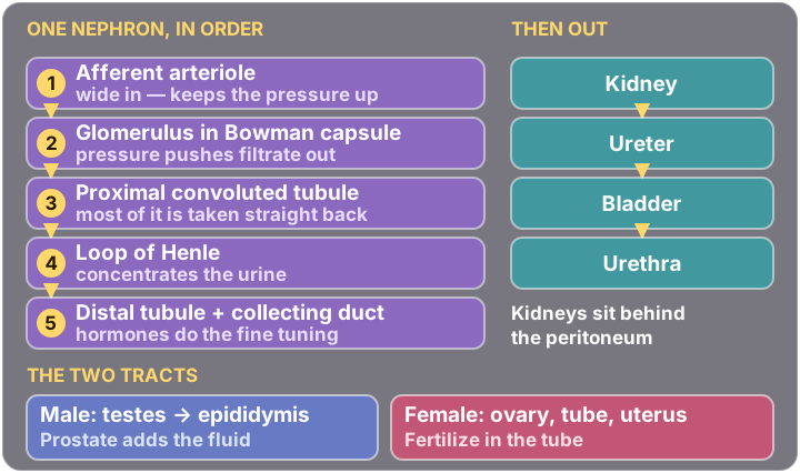
The kidney does not decide what to remove. It removes almost everything, then takes back what it wants. Filter first, reclaim second — which is why the tubule is so long.
Filtration happens in one place: the glomerulus. Everything after it is negotiation.
🥬 The urinary structures
| Structure | Where | Job |
|---|---|---|
| Kidneys | Retroperitoneal — behind the peritoneum | Filter blood, make urine |
| Renal cortex | Outer kidney | Contains the nephrons |
| Renal medulla | Inner kidney | Concentrates urine; holds the pyramids |
| Renal pyramids | In the medulla | Collect and channel urine |
| Ureters | Kidney to bladder | Muscular tubes; move urine down |
| Bladder | Pelvic cavity | Stores urine — has rugae so it can expand |
| Urethra | Bladder to outside | Expels urine |
💧 The nephron, in order
Afferent arteriole brings blood into the glomerulus. Efferent arteriole carries it away.
♂️ Male reproductive structures
| Structure | Where | Job |
|---|---|---|
| Testes | Scrotum | Produce sperm and testosterone |
| Epididymis | On top of the testis | Coiled tube — stores and matures sperm |
| Ductus (vas) deferens | Up from the epididymis | Carries sperm toward the urethra |
| Prostate gland | Below the bladder | Adds alkaline fluid to semen |
| Corpus cavernosum | Penis | Erectile tissue |
| Urethra | Through the penis | Carries both urine and semen |
♀️ Female reproductive structures
| Structure | Where | Job |
|---|---|---|
| Ovaries | Either side of the uterus | Produce eggs and hormones |
| Fallopian tubes | Uterus out toward the ovaries | Carry the egg; fimbriae sweep it in |
| Uterus | Between bladder and rectum | Receives, retains and nourishes the fertilised ovum |
| Cervix | Lower uterus | Connects uterus to vagina |
| Vagina | Cervix to vulva | Birth canal |
| Vulva | External | Labia majora and minora, clitoris |
The uterus has three layers — perimetrium, myometrium (muscle) and endometrium (lining).
Implantation happens in the endometrium.
⚠️ Exam trap
The kidneys are retroperitoneal — behind the peritoneum, not inside the peritoneal cavity. That is why a kidney is approached from the back, and it is a favourite single-word answer.
Take it further🖼️ Renal & Fluid infographics🎯 Quiz me on renal & urinary
BIO 252 · Human Anatomy · fourteen modules, written from your own course study guides and exam review guides. Structure only — the function side lives on the physiology page, and what goes wrong is on pathophysiology.