MI 💔
Myocardial Infarction — the heart muscle DIES
Myocardial Infarction (MI) = the heart muscle DIES — “necrosis” — from lack of oxygen. This happens when there's a blockage of the coronary arteries, the “O₂ tubes” feeding the heart. The blockage is almost always a ruptured atherosclerotic plaque + clot — stage 4 of NG-145. Time = muscle.
📄 Simple Nursing original — opens in Drive →
CAUSE
STEP 1 · THE BLOCKED O₂ TUBEThe coronary arteries feed the heart itself. Block one, and the muscle downstream starves, then dies.
💔 Ischemia → Injury → Infarct: three zones, one blockage
PATHO TIP Necrosis begins within about 20–40 minutes of total occlusion and spreads outward from the inner layer (subendocardium) toward the outer layer over the next several hours. That is why every minute of door-to-balloon time is muscle.
⚠️ Causes — “SODDA”
| Letter | Risk factor | The accurate detail |
|---|---|---|
| S | Stress · Smoking · Stimulants | Caffeine, amphetamines, cocaine — cocaine causes coronary spasm plus clotting (see NG-098) |
| O | Obesity | BMI 25–29.9 = overweight · ≥30 = obesity. Central/abdominal fat carries the most risk. |
| D | Diabetes & hypertension | Older texts: HTN >140/90. 2017 ACC/AHA: HTN starts at ≥130/80; ≥140/90 is stage 2. |
| D | Diet | High cholesterol, animal & trans fats → feeds the plaque (NG-145 & NG-242) |
| A | Age & sex | Risk climbs for men ≥45, women ≥55. Men earlier than women; women catch up after menopause. Black adults carry a higher CVD burden — driven largely by access, chronic stress & social determinants. |
📈 The progression — “CAM”
ACS = unstable angina + NSTEMI + STEMI. The one thing that separates unstable angina from NSTEMI is the troponin.
💔 The MI that isn't a plaque
Most MIs are type 1 — plaque rupture + thrombus. But the muscle can also die when supply and demand split apart:
- 🌀 Coronary spasm — Prinzmetal/variant angina, cocaine (NG-098)
- 📉 Severe hypotension / shock — no pressure, no coronary flow
- 🫀 Sustained tachydysrhythmia — demand outruns supply
- 🩸 Severe anemia or hypoxia — the blood arrives, but empty-handed
- 💉 Coronary embolus or dissection
CLUES
STEP 2 · SPOT ITClassic crushing chest pain is one presentation — and the one that kills quietly is the one that doesn't look like that.
🚨 Signs & symptoms — the classic picture
PAIN: substernal chest pain radiating to the jaw, neck, left arm, mid-back/shoulder, or felt as heartburn (epigastric).
KEY WORDS = PRIORITY “Sudden” · “Crushing” · “Radiating” — if the stem says any of these, treat it as cardiac until proven otherwise.
- 🫁 SOB — “dyspnea,” “labored breathing”
- 🤢 Nausea, vomiting, abdominal pain
- 💦 Diaphoresis — sweating, often cold and drenching
- 🥶 Pale, cool, “dusky” skin
- 😰 Anxiety — classic “impending sense of doom”
- 😵 Dizziness, syncope, palpitations
⭐ ATYPICAL presentations — women, diabetics & older adults
NCLEX TIP A woman or diabetic with unexplained fatigue, nausea and dyspnea gets an EKG — not an antacid. Delay in recognition is a documented reason women have worse MI outcomes.
🆚 STEMI vs NSTEMI vs Unstable angina
| STEMI | NSTEMI | Unstable angina | |
|---|---|---|---|
| Artery | 100% occluded — complete, sustained | Partly / intermittently occluded | Partly occluded, no muscle death yet |
| Depth of damage | Transmural — full thickness of the wall | Subendocardial — inner layer only | None (ischemia only) |
| EKG | ST ELEVATION in ≥2 contiguous leads (new LBBB is treated as an equivalent); Q waves may develop | ST depression and/or T-wave inversion — or a normal EKG | ST depression / T inversion / normal |
| Troponin | ✅ POSITIVE | ✅ POSITIVE | ❌ NEGATIVE — this is the whole difference |
| Treatment urgency | Immediate reperfusion — PCI target ≤90 min door-to-balloon (≤120 min if transferring); fibrinolytic ≤30 min door-to-needle if PCI unavailable | Antithrombotic therapy + risk stratification; cath usually within 24–72 h, sooner if high risk or unstable | Same anti-ischemic therapy, then stratify |
Why the depth matters: a full-thickness (transmural) injury pushes the ST segment up; an inner-layer (subendocardial) injury pushes it down. Same disease, different depth, opposite arrow.
EKG & LABS
STEP 3 · READ THE PROOFEKG first — within 10 minutes of arrival for any chest pain. Then serial troponins. The EKG tells you how fast; the troponin tells you whether.
📈 The four tracings you must recognize on sight
🪦 Pathologic Q wave = the scar
Q waves usually appear hours to days after the infarct and are often permanent — the EKG's tattoo of an old MI.
⏱️ Order of operations
🧪 Cardiac markers — troponin vs CK-MB vs myoglobin
| Marker | Rises | Peaks | Back to normal | Why you'd use it |
|---|---|---|---|---|
| Troponin I / T ⭐ | 3–4 h (high-sensitivity assays: 1–3 h) | 12–24 h | 10–14 days | The diagnostic marker. Most cardiac-specific. Serial draws show the rise and fall. |
| CK-MB | 4–6 h | 12–24 h | 48–72 h | Because it clears fast, a second rise flags RE-infarction while troponin is still elevated. |
| Myoglobin | 1–3 h — earliest | 6–9 h | ~24 h | Fast but not specific — rises with any muscle injury. Mostly historical now. |
| hs-CRP | Inflammation, not injury | Risk stratification only — not an MI diagnostic | ||
SOURCE CHECK A single number like >0.5 ng/mL gets quoted a lot, but troponin cut-offs are assay- and lab-specific — always read the reference range printed on that lab report. And troponin is not the only marker of cardiac injury: it also rises in renal failure, heart failure, PE, sepsis, myocarditis and cardiac contusion. What confirms MI is a rise-and-fall pattern plus a clinical picture, not one number in isolation.
WHICH ARTERY, WHICH WALL
STEP 4 · LOCALIZE ITThe leads with ST elevation tell you which artery is blocked — and which artery tells you which complication to watch for.
🫀 The coronary tree — who feeds what
| Artery | Wall it feeds | Leads with changes | Watch for |
|---|---|---|---|
| LAD left anterior descending |
ANTERIOR wall + SEPTUM + much of the left ventricle | V1 V2 septal · V3 V4 anterior | The “widow-maker.” Biggest infarcts → heart failure, cardiogenic shock, ↓EF, bundle branch blocks |
| LCx left circumflex |
LATERAL wall of the left ventricle (± posterior) | I aVL V5 V6 | Often quieter on the 12-lead — posterior involvement can hide as ST depression in V1–V2 |
| RCA right coronary |
INFERIOR wall, right ventricle, and in most people the SA & AV nodes | II III aVF | Bradycardia & heart blocks · nausea/vomiting · RV infarct → preload dependent |
| Left main | Feeds LAD + LCx = most of the left ventricle | Widespread changes | Catastrophic — very high mortality |
🚨 Inferior MI: the nitro trap
Answer first: Be very cautious with nitroglycerin in an inferior (RCA) MI — up to about half of inferior MIs involve the right ventricle.
- The infarcted RV can't pump forward — it survives on preload
- Nitro drops preload → BP can crash suddenly and severely
- Clues: inferior ST elevation + hypotension + clear lungs + JVD
- Confirm with right-sided leads (V4R)
- Treatment leans on IV fluids to fill the RV
💔 Complications by timeline
- ⚡ Minutes–hours: dysrhythmias — V-fib is the leading cause of early death after MI. This is why she's on a monitor.
- 🫁 Hours–days: heart failure & cardiogenic shock — crackles, ↓BP, ↓urine, cool skin (NG-196)
- 🔊 Days: papillary muscle rupture → sudden new murmur + flash pulmonary edema
- 💥 Days: ventricular free-wall or septal rupture → sudden collapse, tamponade
- 🩹 Days–weeks: pericarditis — sharp pain, better sitting forward, friction rub (Dressler syndrome later)
- 🎈 Weeks: ventricular aneurysm & mural thrombus → embolic stroke risk
CARE
STEP 5 · DURING & AFTERDuring the pain: MONA (with modern asterisks). After: stabilize the clot, rest the heart, and send her home able to protect it.
🚨 DURING — any chest pain: MONA, updated
Why morphine got an asterisk: it still relieves pain, anxiety and preload — but registry data linked morphine to worse outcomes in NSTE-ACS, and it slows absorption of oral antiplatelets (clopidogrel/ticagrelor), delaying their effect. It also masks ongoing ischemic pain, which is one of your best assessment tools. So: reasonable for continuing pain in STEMI, used cautiously and not as a reflex.
Why oxygen got an asterisk: in patients who are not hypoxic, routine O₂ showed no benefit and possible harm — hyperoxia causes coronary vasoconstriction and free-radical injury. Titrate O₂ to a target, don't hand out a nasal cannula by habit.
The real order in practice: Aspirin chewed → Nitro → O₂ only if hypoxic → Morphine only if pain persists. Nurses often say “ANOM” to remember the true sequence.
💊 Nitroglycerin — the hold rules
- 💊 SL 0.4 mg q5 min × 3 max. Pain still there after 3? = MI (injury) — activate EMS/the MI protocol.
- 📉 Nitro drip: hold/stop if SBP <90 or a drop of >30 mmHg from her baseline
- 💥 NEVER with a PDE-5 inhibitor — sildenafil and the other “-afil” drugs — the combination can cause fatal hypotension (avoid within 24 h; ~48 h for tadalafil)
- ⚠️ Caution in RV/inferior MI — preload dependent (see above)
- 🤕 Headache is expected — it means it's working & potent
- 🪑 Change positions slowly — orthostatic hypotension/syncope
- 🌡️ Store in the dark glass bottle, replace per expiration; sit or lie down before taking
🩸 Clot stabilization — heparin
- 🛑 Heparin PREVENTS clot growth — it does NOT dissolve an existing clot
- 💥 Only fibrinolytics ("-ase": alteplase, tenecteplase) actually dissolve clot
- 🧪 Monitor aPTT for IV unfractionated heparin. A commonly taught therapeutic range is 46–70 seconds, but every lab publishes its own — the real target is about 1.5–2.5 × the control value
- 💉 Antidote: protamine sulfate
- 🩸 Monitor platelets — a falling platelet count on heparin suggests HIT
- 📋 Low-molecular-weight heparin (enoxaparin) doesn't need routine aPTT monitoring
SOURCE CHECK A “3× max” is sometimes written for aPTT; the safer teaching is above roughly 2.5× control = bleeding risk. Follow the range printed by your lab.
💊 AFTER the MI — the take-home drug set
| Class | Job | Nursing watch |
|---|---|---|
| A — Antiplatelets aspirin + clopidogrel |
Stop new platelet plugs — especially critical after a stent | Bleeding, bruising, black stools. Many protocols hold if platelets fall to about 50,000/mm³ or less (normal 150–400 K) — follow the order. Antiplatelets are followed by platelet count, not INR or aPTT. |
| C — Cholesterol lowering “-statin” |
Lower LDL and stabilize the plaque cap (NG-145 / NG-242) | Muscle pain/tenderness without injury → report; liver enzymes; no grapefruit; take in the evening |
| B — Beta blockers “-olol” e.g. metoprolol, atenolol |
Lower HR & BP → less O₂ demand, fewer dysrhythmias, less remodeling | Hold parameters: typically HR <60 or SBP <90–100 (per order). Never stop abruptly — rebound tachycardia, HTN and angina |
| A — ACE inhibitors “-pril” then ARBs “-sartan” |
Prevent ventricular remodeling after MI; ACE is first-line, ARB if she can't tolerate ACE | Cough & angioedema = ACE only · monitor K⁺ 3.5–5.0 mEq/L and creatinine (about 0.6–1.2 mg/dL) · avoid in pregnancy · hold for low BP (not low HR) |
| Nitrates isosorbide |
Vasodilate — “a pillow for the heart” | Headache, orthostatic hypotension, never with “-afil” |
| CCBs diltiazem, verapamil, nifedipine |
Vasodilate and (non-dihydropyridines) slow the heart | Diltiazem & verapamil ↓ HR and contractility. “-dipine” drugs are mainly vasodilators and can cause reflex tachycardia — don't expect them to lower the heart rate |
EXAM TIP Beta blockers can mask hypoglycemia in diabetics — they blunt the tremor and tachycardia. Diaphoresis still comes through, so teach her to check glucose rather than wait to “feel low.” Also caution with asthma/COPD (bronchospasm, especially nonselective agents) and in acute decompensated heart failure.
🏠 DISCHARGE — “DRESS” her to go home
NCLEX TIP AVOID NSAIDs (ibuprofen, naproxen) after MI — they increase cardiovascular/clot risk and can blunt aspirin's effect.
⚡ K⁺ & the monitor
- 🧪 Potassium 3.5–5.0 mEq/L — ACE inhibitors and ARBs push it up
- 🥗 Teach: avoid salt substitutes (they're potassium chloride) and don't load up on high-potassium foods without checking
- 📈 High K⁺ = peaked T waves, widening QRS, ST changes → cardiac monitor first
- 💪 Watch for muscle cramps, spasms, weakness
- 📉 Low K⁺ (from diuretics) is just as arrhythmogenic — flattened T, U waves
✅ Nursing priorities in the first hour
- 1️⃣ Assess — pain (OPQRST), vitals, SpO₂, lung sounds, skin
- 2️⃣ 12-lead EKG within 10 min + continuous monitoring
- 3️⃣ Aspirin chewed, IV access ×2, labs including troponin
- 4️⃣ Nitro per protocol with BP before/after every dose
- 5️⃣ Semi-Fowler's, calm environment, keep her at rest — reduce O₂ demand
- 6️⃣ Prepare for reperfusion — cath lab consent, NPO, check bleeding contraindications if fibrinolytics
- 7️⃣ Stay with her — anxiety itself raises catecholamines and O₂ demand
QUICK RECALL
SAY IT OUT LOUD🎯 Cover & check — 8 rapid-fire questions
STUDY SHEETS
FROM YOUR SAVED SET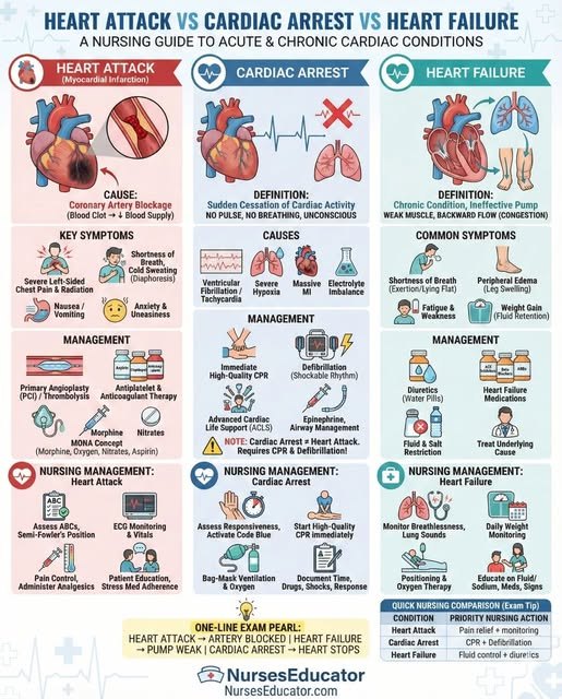
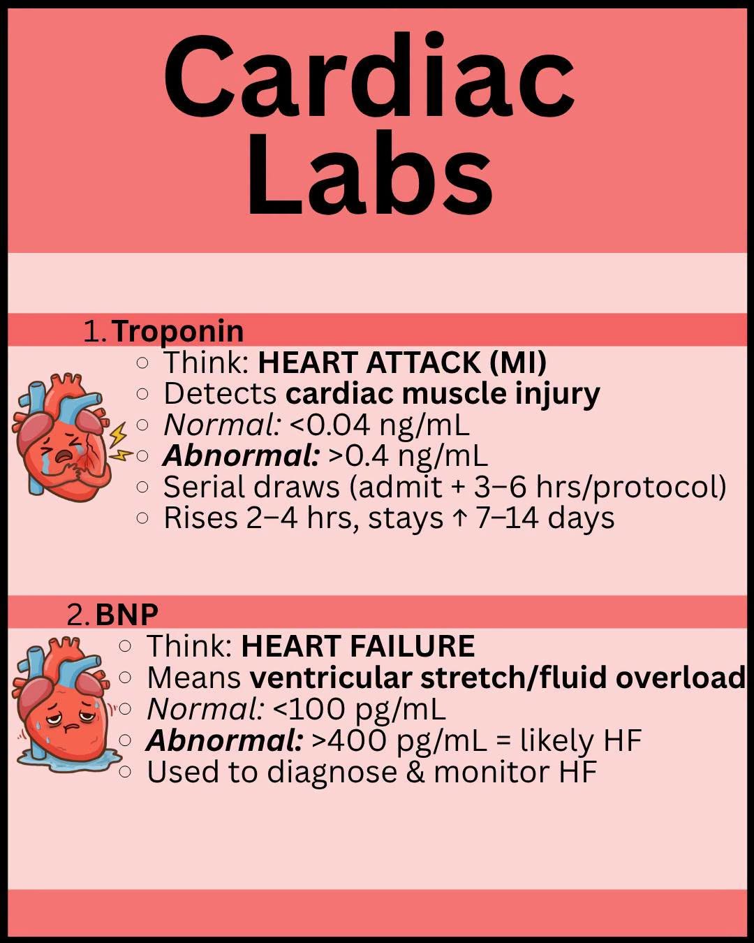
Saved study graphics from your own collection. Each one is someone else’s work — check anything clinical against your course materials before you rely on it.