DVT 🩸
Deep Vein Thrombosis — a CLOT in a deep vein · plus Raynaud's & Buerger's
A clot in a deep vein is not dangerous where it sits — it is dangerous where it GOES. Blood from a leg vein travels to the right heart and straight into the lungs. That is why the calf you find swollen today is a pulmonary embolism tomorrow, and why you never massage or vigorously palpate a calf you suspect of clotting.
📄 Simple Nursing original — opens in Drive →
WHY CLOTS FORM
STEP 1 · VIRCHOW'S TRIADThree conditions make blood clot where it shouldn't. Every risk factor you'll ever be asked about slots into one of them.
🌀 VIRCHOW'S TRIAD — the three-legged stool every DVT stands on
⚠️ Risk factors, sorted by which leg of the triad
| Triad leg | Who's at risk |
|---|---|
| 🧍 STASIS | Post-op & immobile clients · bed rest · casts and splints · paralysis/stroke · long car rides and flights · prolonged sitting · heart failure · obesity · pregnancy (the uterus compresses pelvic veins) |
| 🩹 INJURY | Orthopedic surgery — especially hip and knee replacement · major trauma and fractures · central lines and IV catheters · smoking |
| 🧬 HYPERCOAG | Cancer (and chemotherapy) · dehydration · pregnancy and postpartum · estrogen therapy / oral contraceptives, especially in a smoker over 35 · inherited thrombophilias · sepsis · previous DVT |
The highest-yield combination on the exam: a post-operative orthopedic client who has been in bed and is not yet ambulating. That is all three legs at once.
🫙 Deep vs superficial — why only one of them scares you
- Superficial thrombophlebitis — clot in a surface vein. You can see and feel a red, tender, cord-like vein just under the skin. Uncomfortable, usually treated with warm compresses, elevation and anti-inflammatories.
- DVT — deep vein thrombosis — clot in a deep vein (calf, popliteal, femoral, iliac). You cannot see it. It sits in a high-volume vein with a straight path to the lungs.
- That path is the whole point: leg vein → inferior vena cava → right atrium → right ventricle → pulmonary artery → PE.
SIGNS & ESCALATION
STEP 2 · SPOT ITFour letters for the leg. One line for the lungs. And one thing your hands must never do.
🐄 COWS — the DVT sign set, and the S that changes everything
Also assess: pain on dorsiflexion, a palpable cord, and skin that looks tight and shiny. Do not chart a DVT “ruled out” on findings alone — many DVTs are silent until the PE.
🚫 NEVER massage or vigorously palpate a calf you suspect has a clot
- Why: the thrombus is only loosely attached. Pressure, kneading or forceful dorsiflexion can dislodge it into the circulation, and a venous clot goes straight to the lungs.
- Homans' sign (calf pain on forced dorsiflexion) is no longer used — it is unreliable, it misses most DVTs, and the maneuver itself risks dislodging the clot. Inspect and measure instead.
- Do NOT apply SCDs to a leg with a known acute DVT unless the provider specifically orders it. SCDs are a prevention device.
- Gentle inspection, gentle circumference measurement, and reporting are the assessment. Diagnosis comes from venous duplex ultrasound.
🔬 Confirming it — what actually diagnoses a DVT
- Venous duplex ultrasound — the first-line, non-invasive test. Shows the clot and whether the vein compresses.
- D-dimer — a fibrin breakdown product. A normal D-dimer helps rule OUT a clot; an elevated one does not rule it IN (it rises with surgery, trauma, infection, pregnancy, cancer).
- Wells score — a clinical probability tool that decides whether the client goes straight to ultrasound.
- For suspected PE: CT pulmonary angiography is the standard confirming study. ABG may show hypoxemia with a low CO₂ from blowing off carbon dioxide.
🚨 PE — what it looks like when the clot arrives
- 😮💨 SUDDEN shortness of breath — the hallmark. Abrupt, not gradual.
- 💥 Sharp, pleuritic chest pain — worse on deep breath.
- 💓 Tachycardia and tachypnea; low SpO₂ despite effort.
- 😰 Anxiety, restlessness, a sense of impending doom — take this seriously, it is a real early sign of hypoxia.
- 🩸 Cough, sometimes with blood-streaked sputum (hemoptysis).
- 📉 Hypotension, cyanosis, syncope = massive PE, obstructive shock.
🚨 PE ESCALATION — priority actions in order
Order matters on the exam: when the client is in front of you, do the things that fix oxygenation first (position + O₂), then get help, then gather data. Don't leave the room to page someone.
TREAT & PREVENT
STEP 3 · STOP THE NEXT ONEAnticoagulants don't dissolve the clot — they stop it from growing while the body breaks it down. Prevention is where nursing wins.
💊 Anticoagulant comparison — which lab watches which drug
| Drug | Route & speed | Monitoring | Nursing key points |
|---|---|---|---|
| Heparin unfractionated |
IV drip or SC. Immediate onset — this is the one you start in an emergency. | aPTT (some facilities use anti-Xa). Therapeutic ≈ 1.5–2.5× the control value. Also follow PLATELETS. | Antidote protamine sulfate. Watch for HIT — heparin-induced thrombocytopenia: platelets fall (classically >50% drop) and the client paradoxically clots. Stop the heparin and notify. Never rub SC injection sites. |
| LMWH enoxaparin, dalteparin |
SC, weight-based, more predictable than heparin. | No routine aPTT. Anti-Xa level only in select cases (renal impairment, pregnancy, extremes of weight). Follow platelets and renal function. | Give in the abdomen, about 2 inches from the umbilicus. Do not expel the air bubble, do not aspirate, do not massage the site. Protamine reverses it only partially. |
| Warfarin | PO. Slow — takes several days for full effect, so it is overlapped (“bridged”) with heparin at the start. | PT / INR. Normal INR ≈ 0.8–1.2; therapeutic for VTE/AF is 2.0–3.0. Above range = bleeding risk; below = clot risk. | Antidote vitamin K (phytonadione); major bleeding also gets FFP or prothrombin complex concentrate. Teach consistent vitamin-K intake (green leafy vegetables) — consistency, not avoidance. Many drug/herbal interactions. Contraindicated in pregnancy. |
| DOACs apixaban, rivaroxaban, edoxaban (factor Xa) · dabigatran (direct thrombin) |
PO, fixed dosing, rapid onset. | No routine coagulation monitoring — no INR. Renal function is checked because clearance depends on it. | Reversal: idarucizumab for dabigatran; andexanet alfa for the factor Xa inhibitors. Keep dabigatran in its original bottle and do not crush or open the capsules. Missing doses matters — the protection wears off fast. |
| Thrombolytics alteplase |
IV, reserved for massive PE or a limb-threatening clot. | Continuous monitoring for bleeding of any kind. | Actually dissolves existing clot — and carries the highest bleeding risk of anything on this page. Contraindicated with active bleeding, recent surgery or trauma, and a history of hemorrhagic stroke. |
🩸 Bleeding precautions — the same teaching for every anticoagulant
- 🪥 Soft toothbrush, electric razor, no flossing with force.
- 🚽 Report black tarry stools, pink/red urine, coffee-ground emesis, bleeding gums, nosebleeds, unusual bruising, or a headache/confusion after a fall.
- 💊 No NSAIDs or aspirin unless the provider ordered them; check every OTC and herbal product.
- 🪪 Wear medical-alert identification and tell every dentist and surgeon.
- 🩹 Hold pressure longer after venipunctures and injections; avoid IM injections when possible.
- 🤕 Fall precautions — a head strike on an anticoagulant is an emergency even if the client looks fine.
✅ PREVENTION — the part nursing owns
- 🚶 Early ambulation after surgery — the single most effective prevention.
- 🦵 SCDs / intermittent pneumatic compression and graduated compression (TED) stockings for immobile clients. Check skin under them; make sure they aren't rolled or bunched.
- 💉 Prophylactic anticoagulation — low-dose SC heparin or LMWH for at-risk hospitalized and post-op clients.
- 💧 Hydration — dehydrated blood is thick blood.
- 🦶 Ankle pumps, calf exercises and position changes for anyone on bed rest.
- ❌ No pillows under the knees, no leg crossing, no prolonged sitting — all of them kink the popliteal vein.
🎵 CHANT — living after the clot
Two nuances the exam cares about:
- Elevate the affected leg to promote venous return and reduce swelling — the same rule as every venous problem. SEE NG-161
- About activity during the acute clot: older teaching was strict bed rest to keep the clot from moving. Current practice for most stable clients is early ambulation once therapeutic anticoagulation has been started, because it does not raise PE risk and it reduces pain and swelling. Practice and orders vary — follow the provider's order for your client, and never ambulate a client with a new DVT who has not yet been anticoagulated without checking.
- TEDs and SCDs go on the leg for PREVENTION, and on an affected leg only once the clot is resolved or specifically ordered.
🕸️ When drugs aren't enough: the IVC filter
What it is: a small mesh basket placed in the inferior vena cava to catch clots traveling from the legs before they reach the lungs.
Who gets one: clients who cannot take anticoagulants (active bleeding, recent neurosurgery) or who keep throwing clots despite therapeutic anticoagulation.
What it does NOT do: it does not dissolve clots and it does not prevent new ones from forming — it is a net, not a treatment.
🎨 Also on this card: Raynaud's vs Buerger's
| 🤍 RAYNAUD'S · “R = Ring finger” | 🖤 BUERGER'S · “B = Black fingers & toes” |
|---|---|
| Spasm of small arteries in the fingers (and toes), triggered by COLD and STRESS. | Buerger's disease (thromboangiitis obliterans) — inflammation and clotting of small and medium arteries and veins in the hands and feet. |
| Classic color sequence: WHITE (spasm, no blood) → BLUE (deoxygenated) → RED (blood rushes back). Numbness, tingling, throbbing. | Tissue actually dies: intense pain, claudication of the arch of the foot, cold sensitivity, ulcers, and black gangrenous fingers & toes. |
| Care: keep warm — gloves, warm environment, warm water; avoid cold, caffeine, nicotine; manage stress. Calcium channel blockers are commonly used vasodilators. | Care: absolute, complete tobacco cessation — this is the treatment. Nothing else works if the client keeps smoking. Protect and inspect the extremities; amputation if gangrene. |
| Episodic and reversible. | Progressive and destructive. Strongly associated with tobacco use in younger adults. |
QUICK RECALL
SAY IT OUT LOUD🎯 Cover & check — 8 rapid-fire questions
STUDY SHEETS
FROM YOUR SAVED SET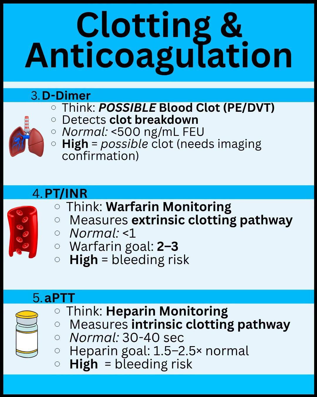
Saved study graphics from your own collection. Each one is someone else’s work — check anything clinical against your course materials before you rely on it.