Nursing Field Notes / Nursing Core · Fundamentals of Nursing
Assessment Locations 📍
Exactly where to put your fingers and your stethoscope
NG-082Nursing CoreADHD-friendly visual edition
Every vital sign from NG-079 has a physical spot on the body where you actually collect it. This page is the landmark map — pulse points, lung auscultation ladder, heart's five listening points, and the abdomen's four quadrants.
⚠️ Never both carotids at onceBilateral pressure can slow heart rate or drop cerebral blood flow.
🫀 "All Pigs Eat Too Much"Aortic → Pulmonic → Erb's → Tricuspid → Mitral, top to bottom.
🩻 IAPP for the bellyInspect → Auscultate → Percuss → Palpate (auscultate BEFORE touching).
💓
PULSE POINTS
STEP 1 · FIND THE BEAT
Same seven sites you saw on NG-079 — here's the exact anatomy and the one you never press on both sides at once.
📍 The seven peripheral pulse points
🧠 "CBR-FPD-T" top to bottom: Carotid, Brachial, Radial, Femoral, Popliteal, Dorsalis pedis, Tibialis (posterior). Walk down the body and you hit every site in order.
⚠️ Caution: carotid pulse
Never palpate both carotid pulses at the same time — bilateral compression can slow the heart rate (vagal response) or reduce blood flow to the brain.
🧠 "One side of the neck at a time — always."
✅ Circulation checks — distal to the pulse
Capillary refill: press nail bed, should return <3 sec
Skin turgor: pinch skin, should return <3 sec
Temperature should be equal bilaterally in all extremities
SEE ALSO NG-190 Head-to-Toe — where these fit in the full exam sequence.
🫁
LUNG AUSCULTATION
STEP 2 · WALK THE LADDER
A systematic zig-zag down the chest, comparing side to side at every stop — never straight down one side then the other.
🪜 The auscultation ladder — anterior & posterior
Start top-right, above the clavicle — listen for a full inhale + exhale
Move to the opposite side, same spot — compare quality, depth, extra sounds
Move down one interspace at a time, comparing side-to-side every stop
Continue through the 6th ICS at the midaxillary line (armpit) — this covers the lower lobes
Flip to posterior: client leans forward, hands in lap to separate the scapulae for a clean listen
🧠 "HF = Heavy Fluid." The lung bases are the best place to catch crackles from fluid overload / heart failure. "A is for Armpit" — the 6th ICS midaxillary line is right at the armpit and picks up the lower lobes.
🫱 Tactile fremitus
Use the ulnar and palmar surfaces of your hands against the chest wall while the client says "ninety-nine" — vibration should feel equal bilaterally.
Correct client language for shortness of breath: "It's hard for me to breathe and I feel short winded all the time."
Dys-PNEA = Difficult breathing — a PRIORITY symptom.
🚨 Priority interventions — "breathing problems"
Ensure the airway is open
Monitor O2 saturation
Place client in High Fowler's
Evaluate effectiveness of respiratory effort after intervening
🧠 Most important objective data confirming hypoxia: ABGs (arterial blood gases) — not just the SpO2 number.
👀 Cyanosis assessment sites
Bluish discoloration from hypoxia is checked at:
Nailbeds — peripheral cyanosis
Conjunctiva — central/mucosal cyanosis, more reliable in darker skin tones
Earlobes
🧠 Also watch for: clubbing of the fingernail beds (chronic hypoxia), vertigo/dizziness, and increased pulse rate as signs of longer-term hypoxia.
🫀
HEART & ABDOMEN
STEP 3 · THE OTHER TWO STOPS
Five points for the heart, four quadrants for the belly — and one order you can't mix up.
🫀 Heart auscultation — 5 points, "All Pigs Eat Too Much"
🧠 "All Pigs Eat Too Much" — Aortic, Pulmonic, Erb's point, Tricuspid, Mitral, top to bottom down the left sternal border, ending at the apex (mitral/apical — the same spot you count the apical pulse from NG-079).
🩻 Bowel sounds — four quadrants
Listen for sounds in all four quadrants. If absent, listen for a full 5 minutes before charting "absent."
🧠 Chart as active, hyperactive, or hypoactive — never just "present."
🚨 The order you can't mix up: IAPP
1️⃣ Inspect
▼
2️⃣ Auscultate
▼
3️⃣ Percuss
▼
4️⃣ Palpate
The abdomen is the ONE exception to the usual inspect→palpate→percuss→auscultate order — palpating or percussing first can artificially stimulate bowel sounds and give a false reading.
🧠 "Look, Listen, THEN Touch" for the belly — auscultate before you ever press on it.
🫃 Normal abdominal shape
Belly should be flat, non-pulsating, and even/symmetric. Ask about pain with palpation — a rigid, board-like abdomen with fever and tachycardia is peritonitis until proven otherwise and is a priority to report.
SEE ALSO NG-190 Head-to-Toe Assessment — where heart, lung, and abdominal auscultation fit into the full systematic exam sequence.
⚡
QUICK RECALL
SAY IT OUT LOUD
💓 7 pulse pointsnever both carotids at once
🫁 6th ICS midaxillarylowest stop on the ladder = lower lobes
🫀 APE To ManAortic → Pulmonic → Erb's → Tricuspid → Mitral
🩻 Auscultate before palpateonly true for the abdomen
🎯 Cover & check — 4 rapid-fire questions
Q1: Why should you never palpate both carotid pulses at once?
Bilateral pressure can slow the heart rate via a vagal response or reduce cerebral blood flow.
Q2: Where is the best place to auscultate for fluid overload in the lungs?
The posterior lung bases — listen for crackles ("HF = Heavy Fluid").
Q3: What's the correct order of abdominal assessment, and why is it different from other systems?
Inspect, Auscultate, Percuss, Palpate — auscultate before touching so you don't artificially stimulate bowel sounds.
Q4: List the five heart auscultation points in order.
Aortic (2nd R ICS) → Pulmonic (2nd L ICS) → Erb's point (3rd L ICS) → Tricuspid (4th L ICS) → Mitral (5th L ICS, midclavicular) — "All Pigs Eat Too Much."
📌
STUDY SHEETS
FROM YOUR SAVED SET
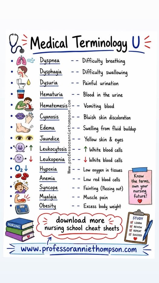
The terms that turn up in every question stem — dyspnea, dysphagia, dysuria, hematuria, hematemesis, cyanosis, edema, jaundice and the rest. — swipe it sideways if it is cut off, or tap to open it full size.
Saved study graphics from your own collection. Each one is someone else’s work — check anything clinical against your course materials before you rely on it.