Bleeding anywhere inside the GI tract — the whole job is figuring out WHERE. Split the tube at the ligament of Treitz: bleed above it is an upper GI bleed, bleed below it is a lower GI bleed. Enough blood loss fast enough → hypovolemic (hemorrhagic) shock.
🩸 Coffee grounds & tar = UPHematemesis / melena = upper GI bleed.
🍷 Bright red = DOWNHematochezia = lower GI bleed.
🧪 Hgb < 7 = transfuse"Hemoglobin less than 7 = heaven" (send blood).
❌ No NGT + varicesNever lavage a client with known esophageal varices.
🧨
CAUSE
STEP 1 · FIND THE TUBE, PICK A SIDE
One landmark splits the whole GI tract into "upper" and "lower" — everything else follows from that split.
🗺️ The ligament of Treitz is the dividing line
DEFINITION A GI bleed is bleeding anywhere inside the GI tract, classified as upper or lower — one of the biggest jobs is figuring out the exact location because it changes every intervention that follows (NGT? scope type? drug?).
🧠 "Treitz splits the tube." Above the ligament of Treitz = upper (mouth-side). Below it = lower (anus-side). Everything about a GI bleed — the symptom, the scope, the NGT rule — depends on which side.
⚠️ Upper GI bleed — causes
🔥 Gastritis
🚪 GERD / esophagitis
🕳️ Peptic ulcer disease — most common cause
🩸 Esophageal varices — from portal hypertension / cirrhosis, can rupture & exsanguinate fast
🤮 Mallory-Weiss tear — forceful vomiting/retching
🧠 "GGPV" — Gastritis · GERD · Peptic ulcer · Varices. The V is the scary one — varices bleed the fastest and hardest.
🚨 RED FLAG — call the provider now (hemorrhagic shock)
Any GI bleed can outrun the body's ability to compensate. These signs mean the client is decompensating — report immediately:
📉Hypotensionfalling BP
💓TachycardiaHR > 100
🥶Cool, clammydiaphoretic
😶Pale skin
😵Dizzy, fatigue
🧪Hgb < 7transfusion trigger
🧠 "Cool, pale, fast, low" — cool clammy skin + pale + fast HR + low BP is the classic hemorrhagic shock picture. Don't wait for BP to crash — tachycardia is the FIRST compensatory clue.
🔎
CLUES
STEP 2 · READ THE BLOOD
What color, and which end — that's the whole exam question.
Hematochezia — bright red blood per rectum (fresh, undigested)
BUN:Cr ratio
Often elevated — digested blood protein is absorbed and broken down to urea
Usually normal
Scope used
EGD (esophagogastroduodenoscopy)
Colonoscopy
🧠 "Coffee grounds and tar are OLD — bright red is FRESH." The higher up the bleed, the more time acid & digestion have to darken it before it comes out.
🖼️ Coffee-ground emesis vs. bright red stool
📉 Hypovolemic shock cascade
🩸 Blood volume drops
▼
💓 Compensatory tachycardia
▼
📉 Hypotension — decompensating
▼
🥶 Cool, clammy, pale, dizzy, confused
🧠 Tachycardia shows up before BP drops — it's the body compensating. Don't wait for hypotension to call it a bleed.
🧪 Labs — trend the H&H
Low CBC labs = low H/H (hemoglobin & hematocrit).
critical
low
normal
Adult reference (varies by lab): Hgb M 14–18 / F 12–16 g/dL. Hgb < 7 g/dL = transfusion trigger.
🧠 "Hemoglobin less than 7 = heaven" — rhymes on purpose. Below 7, send blood.
🧫 Diagnostics — Guaiac Fecal Occult Blood Test (gFOBT)
1
🧤 Gather supplies, wash hands, don non-sterile gloves
2
📦 Open card, apply stool sample to both boxes on the slide
3
🧴 Open the back, apply 2 drops developing solution, wait 30–60 sec
4
📝 Document results — blue = positive for occult blood
Definitive location & source is found by EGD (upper) or colonoscopy (lower) — the scope both diagnoses and treats.
🩺
CARE
STEP 3 · STOP THE BLEED, REPLACE THE VOLUME
Stabilize first, scope second, feed last.
✅ Priority nursing interventions
1
Lower the head of the bed — improves perfusion to vital organs
2
IV normal saline — 2 large-bore IVs, stabilizes blood pressure
3
Oxygen
4
Blood transfusion — when Hgb < 7, type & crossmatch PRBCs
🧠 "LOB" — Lower HOB · Oxygen & IV fluids · Blood if Hgb < 7. Also: keep the client NPO in case of emergency endoscopy.
❌ NGT lavage — upper GI only, one huge exception
Nasogastric lavage (NGT) can be used to assess/clear an upper GI bleed.
NEVER insert an NGT if esophageal varices are known or suspected — the tube can rupture the fragile varix and cause catastrophic bleeding.
🧠 "Varices + tube = trouble." If the chart says cirrhosis or varices, the NGT answer on the exam is always "no."
💊 IV PPI (e.g., pantoprazole) — reduces acid, protects a healing ulcer
🩸 Blood products — PRBCs, FFP, platelets as ordered
🧠 Octreotide = "shrink the plumbing." It squeezes down splanchnic vessels so varices bleed less.
🔪 Surgery / procedures — find it & fix it
🔭 Endoscopy (EGD) — upper GI: cauterize, clip, or band a bleeding varix/ulcer
🔭 Colonoscopy — lower GI: locate & treat the source
🩹 Angiography / embolization if bleeding can't be localized or controlled by scope
🧠 "Scope up = upper, scope down = lower." EGD goes in the mouth; colonoscopy goes in the rectum.
🍽️ After the "-scopy" — diet progression
Client stays NPO before the procedure, then advances slowly once bleeding is controlled and gag reflex has returned:
🍎Apple juiceclear liquid
🍲Broth soupclear liquid
🍵Unsweetened teaclear liquid
Advance diet as tolerated — clear liquids → full liquids → soft → regular, watching for return of nausea, pain, or bleeding.
⚡
QUICK RECALL
SAY IT OUT LOUD
🩸 Coffee grounds / melena= upper GI bleed
🍷 Hematochezia= lower GI bleed, bright red
🧪 Hgb < 7= transfuse (heaven)
❌ Varices= never NGT lavage
🎯 Cover & check — 4 rapid-fire questions
Q1: Client has coffee-ground emesis and black tarry stools. Upper or lower GI bleed?
Upper — hematemesis/coffee-ground emesis and melena are both upper GI bleed clues.
Q2: What lab value triggers a blood transfusion?
Hemoglobin less than 7 g/dL — "Hgb less than 7 = heaven."
Q3: Client with known esophageal varices needs gastric lavage. What do you do?
Do NOT insert an NGT — it can rupture the varix. Notify the provider instead.
Q4: HR 128, BP 84/52, skin cool and clammy. What's happening and what's priority?
Hypovolemic (hemorrhagic) shock. Priority: lower HOB, 2 large-bore IVs with normal saline, oxygen, notify provider, prepare for possible transfusion.
📌
STUDY SHEETS
FROM YOUR SAVED SET
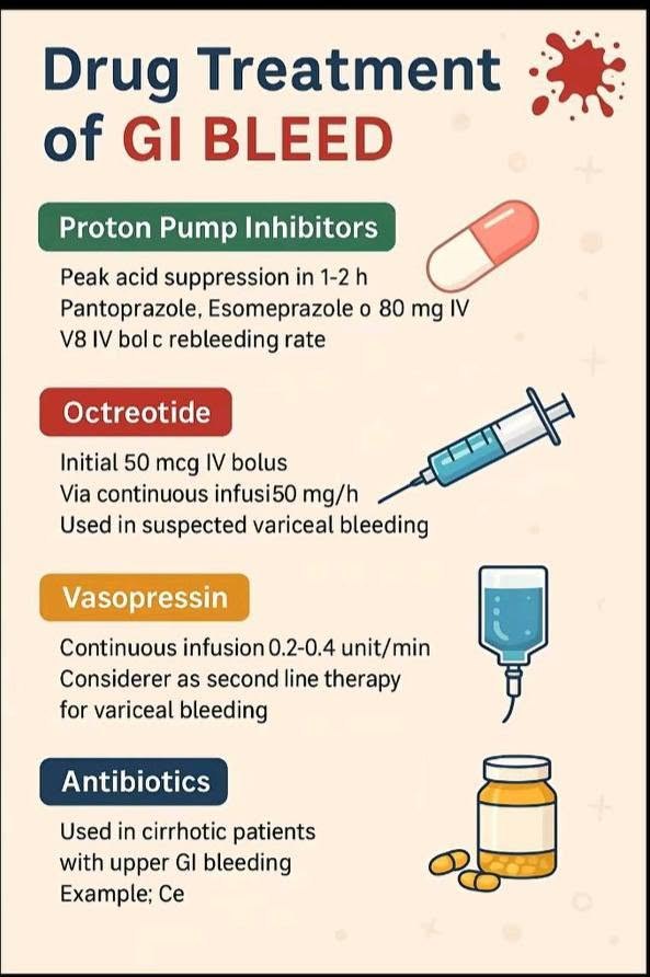
The four drug classes for a GI bleed side by side — PPI, octreotide, vasopressin and antibiotics — with the doses and which bleed each one is for. — swipe it sideways if it is cut off, or tap to open it full size.
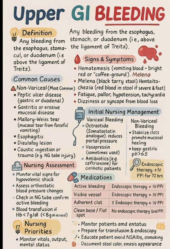
Upper GI bleeding in full — above the ligament of Treitz, variceal versus non-variceal, and the table of what each endoscopic finding gets. — swipe it sideways if it is cut off, or tap to open it full size.
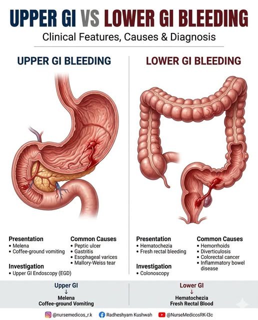
Upper versus lower GI bleed at a glance. Upper: melena and coffee-ground vomiting, EGD. Lower: hematochezia and fresh rectal blood, colonoscopy. — swipe it sideways if it is cut off, or tap to open it full size.
Saved study graphics from your own collection. Each one is someone else’s work — check anything clinical against your course materials before you rely on it.