Degenerative Joint Disease & Total Knee Replacement
NG-168MusculoskeletalADHD-friendly visual edition
Osteoarthritis is "wear and tear," not autoimmune — cartilage wears down until it's bone-on-bone, and pain gets worse with activity, better with rest. When conservative care fails, a total knee arthroplasty (TKR/TKA) replaces the joint — and post-op nursing care is a heavily tested skill of its own.
🦴 OA = bone-on-boneCartilage degenerates from wear; pain worsens with activity, eases with rest.
⏱️ Stiffness <30 min = OARA's morning stiffness lasts >30–60 min and eases with movement — opposite pattern.
🦵 No pillow under the kneePromotes flexion contracture after TKR — keep the leg extended.
🩸 Heparin + warfarin togetherStarted same day — heparin acts fast, warfarin takes days to catch up.
🦴
OA PATHOPHYSIOLOGY
STEP 1 · WEAR & TEAR
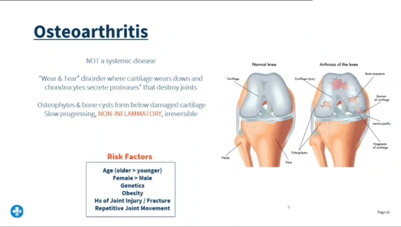
One mechanism explains every OA finding: the cartilage cushion wears away until bone grinds on bone.
From your lecture.Not a systemic disease and non-inflammatory — a wear-and-tear disorder that is slow and irreversible. Risk: older age, female, genetics, obesity, prior joint injury, repetitive movement.
🦴 Healthy cartilage vs. osteoarthritic joint
Progressive degeneration of the protective cartilage cushion results in bone-on-bone rubbing and massive pain.
🧠 "OA-Ouch pain." The cushion wore out — think of a worn-out shoe sole slapping the pavement.
⚠️ Causes & risk factors
Age — cumulative wear over decades.
Obesity — extra load on weight-bearing joints.
Repetitive stress/overuse on the joints (occupational, athletic).
Prior joint trauma or injury.
🧠 Load + time = OA. Any factor that adds mechanical stress or years of use raises risk.
🔎 Signs & symptoms
Crepitus — a "crunch" sound/feel with joint movement.
Pain worse with activity, relieved by rest.
Usually affects one joint at a time — typically a large, weight-bearing joint (knee, hip) — asymmetric.
Heberden's nodes (DIP joints) and Bouchard's nodes (PIP joints) — bony hand nodules.
🧠 "Heberden's is Higher up the finger." Heberden's = distal (DIP, closer to nail); Bouchard's = proximal (PIP, closer to hand) — say them in that finger order to keep them straight.
✋ Heberden's vs. Bouchard's nodes
🧠 Use the fingertip, not the alphabet. Heberden's is closest to the nail (H = high, at the tip = DIP); Bouchard's sits at the middle knuckle (PIP).
🔎
OA vs. RA
STEP 2 · TELL THEM APART
These two get confused on every exam — one table settles it. See the Rheumatoid Arthritis page (NG-163) for RA's full autoimmune picture.
⚖️ Osteoarthritis vs. Rheumatoid Arthritis
Osteoarthritis (OA)
Rheumatoid Arthritis (RA)
Degenerative — mechanical wear, NOT autoimmune
Autoimmune — systemic inflammatory disease
Morning stiffness <30 minutes
Morning stiffness >30–60 minutes
Pain worse with activity, better with rest
Pain/stiffness worse with rest, improves with movement
Asymmetric — one joint, usually large/weight-bearing (knee, hip)
Symmetric — small joints of hands/wrists on both sides
No systemic symptoms; local joint disease only
Systemic — fatigue, low-grade fever, malaise, weight loss
🧠 "OA-Ouch pain, RA-Rude pain." OA hurts more the more you use it (ouch, overuse). RA is rude first thing in the morning and calms down once you get moving.
🚨 SATA-style NCLEX manifestations of OA
✔️ Joint pain that diminishes after rest
✔️ Joint pain that intensifies with activity
✔️ Node formation (Heberden's/Bouchard's)
✔️ Affects one joint, usually large
🧠 Select-all-that-apply questions test whether you know the OA pattern is activity-triggered, not inflammatory-triggered.
💊 Medication risk factors to recognize
NSAIDs — first-line for pain/inflammation, but GI bleed risk.
Corticosteroids ("-sone," e.g. prednisone) — can worsen cartilage breakdown and mask infection with long-term use.
Glucosamine — supplement some clients use for joint support; evidence is mixed, but it's a commonly tested item.
🧠 "NSAIDS, glucosamine, prednisone" = the most-tested trio for OA symptom management.
🔪
TKR: PRE/POST-OP CARE
STEP 3 · PROTECT THE NEW JOINT
A total knee arthroplasty replaces the joint surface — nursing care afterward focuses on clots, motion, and neurovascular status.
🩹 Total knee arthroplasty (TKA/TKR) — the basics
A surgical procedure to replace the knee joint when conservative management of severe OA (or RA) fails. Also a common surgical intervention for RA-damaged joints.
🧠 The joint surfaces are replaced with metal/plastic components — the whole leg isn't hollowed out, just the worn cartilage/bone ends.
🚨 Neurovascular checks — the 6 P's
Pain (out of proportion)
Pallor
Pulselessness
Paresthesia
Paralysis
Poikilothermia (coolness)
🧠 Same 6 P's used for any extremity at risk of compromised circulation — see the Fracture (NG-056) page, where these first appear for compartment syndrome screening.
✅ Post-op priorities
1
Flex the foot every hour while awake — ankle pumps to prevent DVT.
2
Early weight-bearing/ambulation as ordered, PT-directed.
3
CPM machine (continuous passive motion) to gradually increase knee flexion.
4
Ice, elevation of the whole leg (not just under the knee), and pain control to enable participation in PT.
🧠 "Flex it hourly, move it early." Motion is medicine after a TKR — both for the joint and for clot prevention.
❌ Never place a pillow under the operative knee
Never place a support pillow directly under the new operative knee — it holds the joint in flexion for prolonged periods, which promotes a flexion contracture and blocks the surgical goal of full extension/flexion range.
🧠 Straight leg, straight recovery. A pillow under the knee feels comfortable but locks the joint in the wrong position — keep the leg extended when resting, per care team positioning orders.
🩸 Clot prevention: heparin bridges to warfarin
Heparin and warfarin are often started at the same time in the hospital, because heparin works fast (immediate anticoagulation) while warfarin has a slow onset — it takes several days to reach a therapeutic effect. The two overlap until warfarin catches up, then heparin is stopped.
🧠 "Heparin sprints, warfarin walks." Start together, bridge the gap, then drop the sprinter once the walker (warfarin/INR) reaches target.
⚡
QUICK RECALL
SAY IT OUT LOUD
🦴 OA = wear & tearPain worse with activity, better with rest; one joint at a time.
⏱️ <30 min stiffnessOA's morning stiffness is brief — RA's lasts >30–60 min.
🦵 No pillow under the kneePrevents flexion contracture after TKR.
🩸 Heparin + warfarinStarted together — fast bridge to slow, long-term anticoagulation.
🎯 Cover & check — 4 rapid-fire questions
Q1: How does OA pain differ from RA pain in timing?
OA pain worsens with activity and improves with rest, with morning stiffness under 30 minutes. RA is the opposite — worse with rest/morning, improves with movement, and morning stiffness lasts over 30–60 minutes.
Q2: A nurse is teaching a client after TKR. What foot/ankle instruction should be included?
Flex the foot every hour while awake (ankle pumps) to promote circulation and reduce DVT risk.
Q3: Why should a pillow never be placed directly under the operative knee after TKR?
It holds the knee in prolonged flexion, which can cause a flexion contracture and prevent the client from reaching full extension.
Q4: Why are heparin and warfarin often started on the same day after a TKR?
Heparin acts immediately but warfarin takes several days to reach therapeutic effect — the two overlap so the client stays protected while warfarin "catches up," then heparin is discontinued.