Nursing Field Notes / Musculoskeletal · Fracture & Trauma Care Series · Page 1 of 4
Fracture 🦴
Bone Breaks, Neurovascular Checks & Emergency Stabilization
NG-056MusculoskeletalADHD-friendly visual edition
A fracture is any break in the continuity of a bone — from a hairline crack to a shattered shaft. The nurse's job isn't the x-ray, it's everything around the bone: is the skin open, is the limb still getting blood flow and nerve signal, and is there a life threat (pelvic bleed, spinal shock, skull fracture) hiding behind the obvious injury? This page builds the 6 P's — the neurovascular assessment used on every single page in this series.
🚨 Splint before you moveImmobilize the joint above & below the break first — always.
🦴
TYPES
STEP 1 · NAME THE BREAK
Every fracture question starts by naming what kind of break it is — the name usually tells you the risk.
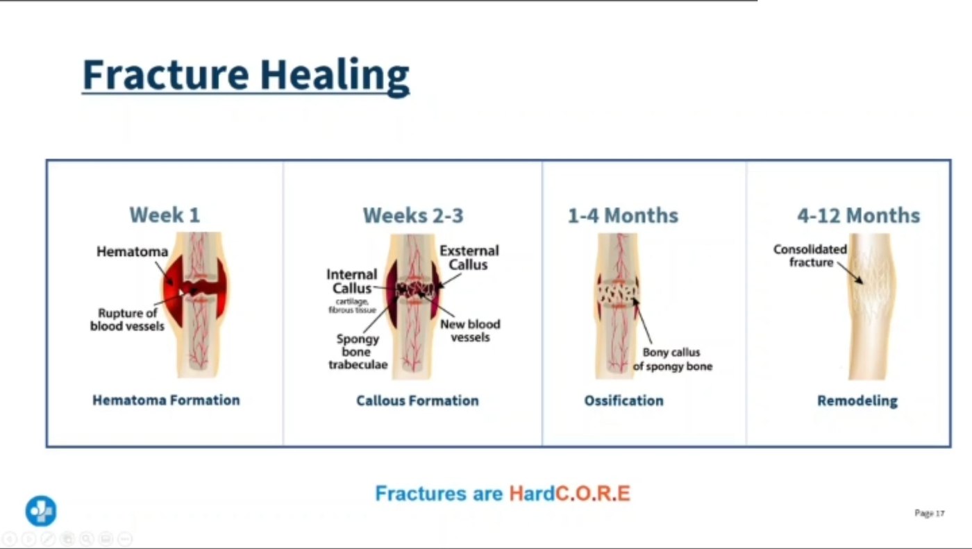
From your lecture. Four stages, and the timescale is the examinable part: hematoma → callus → ossification → remodeling, week 1 out to 12 months.
🩻 Six fracture patterns you must recognize on sight
🧠 “COGS-C” — Closed · Open · Greenstick · Spiral · oblique(Cut diagonal) · Comminuted/compression. Whatever the name — always ask “is the skin open?” first, because that changes everything about infection risk.
🚪 Closed vs Open — the one question that changes care
Closed
Open “compound”
Skin intact — bone does not break the skin surface
Skin surface broken — bone visible or was exposed
Lower infection risk
High infection risk — ask about last tetanus vaccine
Cover, splint, x-ray
Sterile dressing, never push the bone back in, IV antibiotics often started
🧠 HESI trap: open leg fracture → the nurse should ask “when was your last tetanus shot?” — broken skin + soil/debris exposure = tetanus risk.
⚠️ Risk factors — what sets a fracture up
🛏️ Prolonged bed rest — disuse osteoporosis, muscle atrophy & loss of tone
🦴 Osteoporosis — weak, porous bone (see the Osteoporosis page in this library)
💊 Steroids “-sone” — prednisone, dexamethasone, hydrocortisone, fludrocortisone — long-term use thins bone
💥 Trauma — falls, MVC, sports
🧠 Kaplan SATA: long-term bed rest effect the nurse should worry about most → loss of muscle tone and disuse atrophy (select-all-that-apply style question).
🖐️
ASSESS
STEP 2 · THE 6 P's
This is the neurovascular check you will run on every injured or casted limb — memorize it here, it repeats on every page in this series.
🖐️ THE 6 P's — neurovascular assessment of an injured limb
🧠 “Early vs late.”Paresthesia (tingling) shows up first — that's your window to intervene. Pulselessness and paralysis are late signs — by the time you see those, tissue damage may already be underway. Never wait for all 6 P's — one abnormal P = notify the HCP.
🩸 Pelvic fracture — the priority finding you must catch
💥 Pelvic fracture — large vessels run right through the pelvis
▼
🩸 Internal bleeding into the pelvic cavity (can be liters, hidden)
Also watch for: crepitus (grating sensation/sound of bone ends rubbing) and muscle spasm around the fracture site.
🧠 Saunders trap: "Which is the most serious finding for a pelvic fracture?" → hypotension, tachycardia, and hematuria — signs of hemorrhagic shock and bladder/urethral injury, not just local pain.
🦵 Hip fracture — the classic exam picture
📏 Shortening of the affected leg
🔄 Affected leg often externally rotated
😖 Muscle spasm around the hip
🟣 Ecchymosis (bruising) over thigh/hip
⚡ Groin & hip pain that worsens with weight bearing
🧠 Hip fracture = “short & turned out.” A leg that looks shorter and rotated outward, in an older adult who fell, is a hip fracture until proven otherwise.
🧠 Skull, spine & mandible — trauma above the neck
Basilar skull fracture: clear fluid from nose/ear (CSF rhinorrhea/otorrhea) — never suction or pack the nose, test for a "halo" ring on gauze.
Spinal fracture at T6 or higher → neurogenic shock:hypotension + bradycardia + skin that stays pink & dry (loss of sympathetic tone, not hypovolemia).
Mandibular fracture: bleeding/drooling from the mouth → suction the mouth, protect the airway first.
🧠 “Bradycardia + hypotension + warm dry skin” = neurogenic shock, NOT hypovolemic shock (which is tachycardic, cool, clammy). Easy to mix up on an exam.
🩺
CARE
STEP 3 · STABILIZE FIRST
Immobilize before you move anything — then reassess the 6 P's before and after every intervention.
🚑 Emergency stabilization — in order
1
ABCs first — treat life threats (airway, breathing, hemorrhage) before the limb itself
2
Assess the 6 P's distal to the injury before touching it
3
Splint the joint above AND below the fracture before any movement
4
Reassess the 6 P's immediately after splinting — a tight splint can cause the exact injury it prevents
5
Elevate & ice the limb to reduce swelling
🧠 “Check, splint, check again.” Neurovascular status is assessed before AND after every splint, cast, or traction device is applied — that repeated check is the single most tested nursing action in this whole unit.
⏳ Fracture healing timeline — what's happening under the cast
🧠 “Clot → Soft → Hard → Remodel.” A soft callus (week 1–2) will bend or re-break under stress — that's exactly why a fresh cast means no weight-bearing yet, even though the limb is immobilized. Full remodeling can take up to two years.
🚨 Open fracture — do this, never that
✅ Cover exposed bone with a sterile dressing
✅ Immobilize/splint as-is
✅ Confirm tetanus vaccination status
❌ Never push exposed bone back under the skin
❌ Never irrigate or clean the wound aggressively in the field
🧠 Cover it, don't cover it up. The bone stays where it is — the HCP reduces it in a controlled, sterile setting.
🧊 Pain, swelling & the compartment syndrome watch
Fracture pain classically includes bruising, swelling, muscle spasm, and crepitus (grating). Elevate the limb, apply ice, and treat pain proactively.
Pain that keeps climbing despite analgesia — especially with passive stretch — is not "just a bad fracture." It is the first sign of compartment syndrome, covered in full on the Cast Care page in this series.
🧠 If pain doesn't match the picture — always think compartment syndrome, not "needs more pain meds."
➡️ Where this story goes next
Fracture care doesn't stop at the ED. This series follows the same patient forward:
🛏️ NG-058 Buck's Traction & postoperative musculoskeletal care
🫁 NG-061 Fat embolism & osteomyelitis — the two big fracture complications
🦿 NG-063 Cast care — teaching the 6 P's back to the patient at home
🧠 The 6 P's you learned above are the exact same checklist used for traction pin sites, casted limbs, and post-op neurovascular checks — one skill, reused everywhere in orthopedics.
⚡
QUICK RECALL
SAY IT OUT LOUD
🦴 Open = tetanus checkclosed = skin intact, open = skin broken + infection risk
Q1: What's the difference between a closed and an open fracture?
Closed: skin intact. Open/compound: bone breaks through the skin — high infection risk, check tetanus status, never push the bone back in.
Q2: Name all 6 P's of neurovascular assessment.
Pain, Pallor, Pulselessness, Paresthesia, Paralysis, Poikilothermia (temperature). Paresthesia is early; pulselessness and paralysis are late/ominous.
Q3: A client with a pelvic fracture is hypotensive, tachycardic, and has hematuria. What's happening?
Internal bleeding from the pelvic fracture — report immediately, this is the most serious finding to catch.
Q4: When do you assess the 6 P's on a splinted or casted limb?
Before AND after applying any splint, cast, or traction device — and regularly afterward.
📌
STUDY SHEETS
FROM YOUR SAVED SET
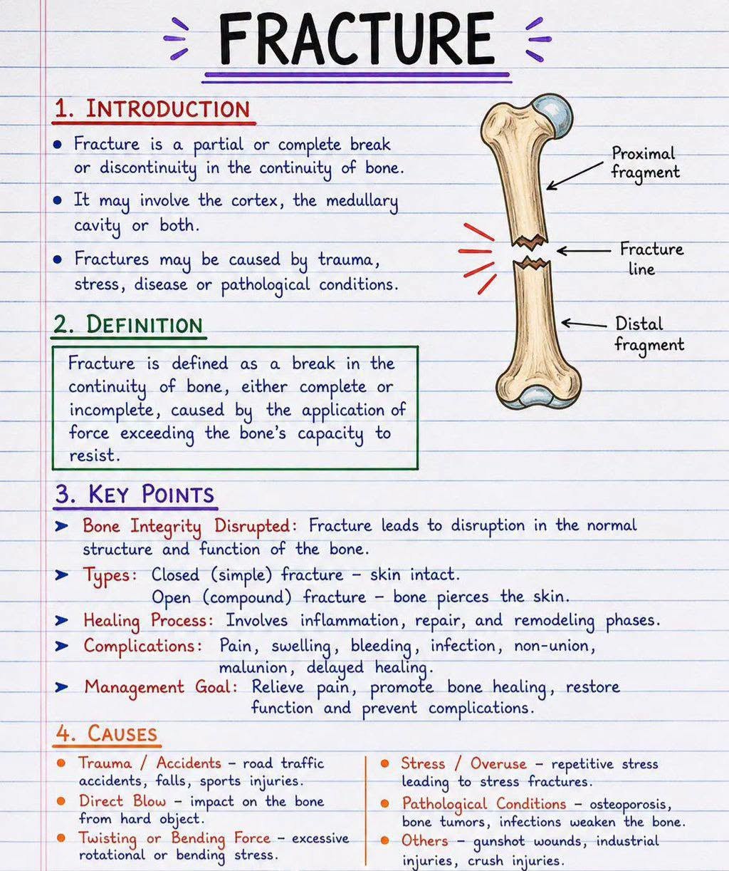
Part 1 — what a fracture is, the key points, and the causes: trauma, direct blow, twisting, repetitive stress and pathological conditions. — swipe it sideways if it is cut off, or tap to open it full size.
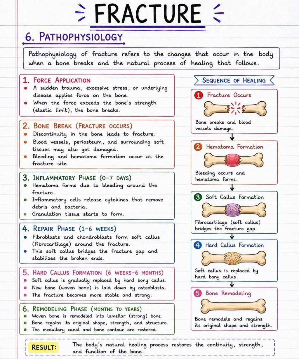
Part 2 — the five stages of bone healing in order: hematoma, inflammation, soft callus, hard callus, remodeling, with the weeks each takes. — swipe it sideways if it is cut off, or tap to open it full size.
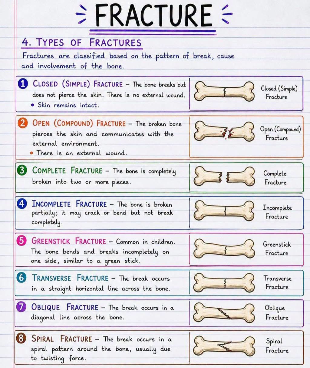
Part 3 — the eight fracture types drawn: closed, open, complete, incomplete, greenstick, transverse, oblique and spiral. — swipe it sideways if it is cut off, or tap to open it full size.
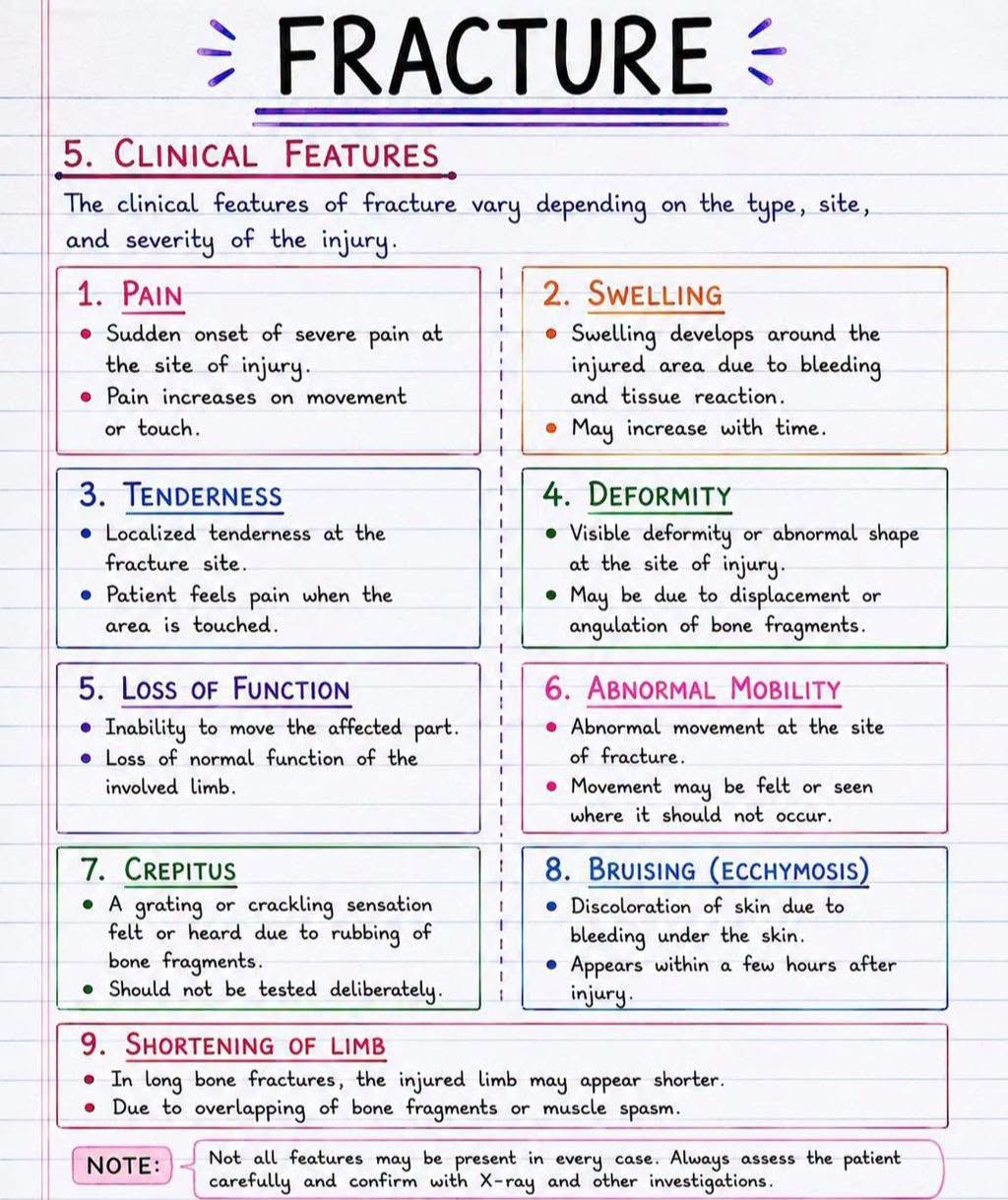
Part 4 — the nine clinical features, including crepitus (never test for it deliberately) and shortening of the limb. — swipe it sideways if it is cut off, or tap to open it full size.
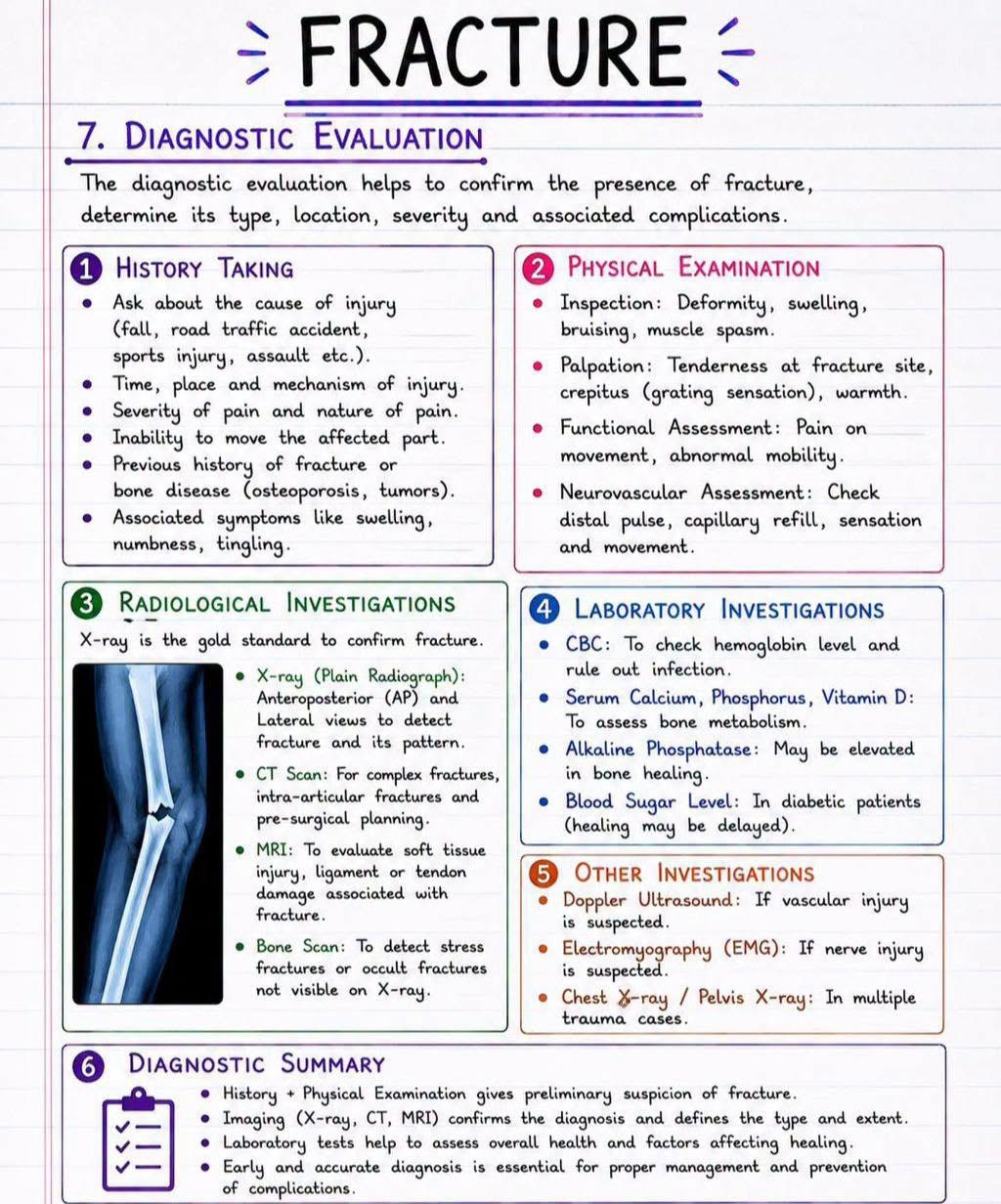
Part 5 — history, physical examination and the imaging: X-ray is the gold standard, CT for complex, MRI for soft tissue, bone scan for stress fractures. — swipe it sideways if it is cut off, or tap to open it full size.
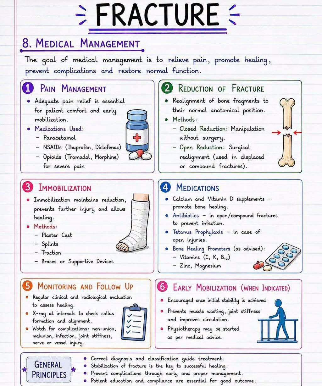
Part 6 — pain control, closed versus open reduction, immobilization, monitoring, and early mobilization. — swipe it sideways if it is cut off, or tap to open it full size.
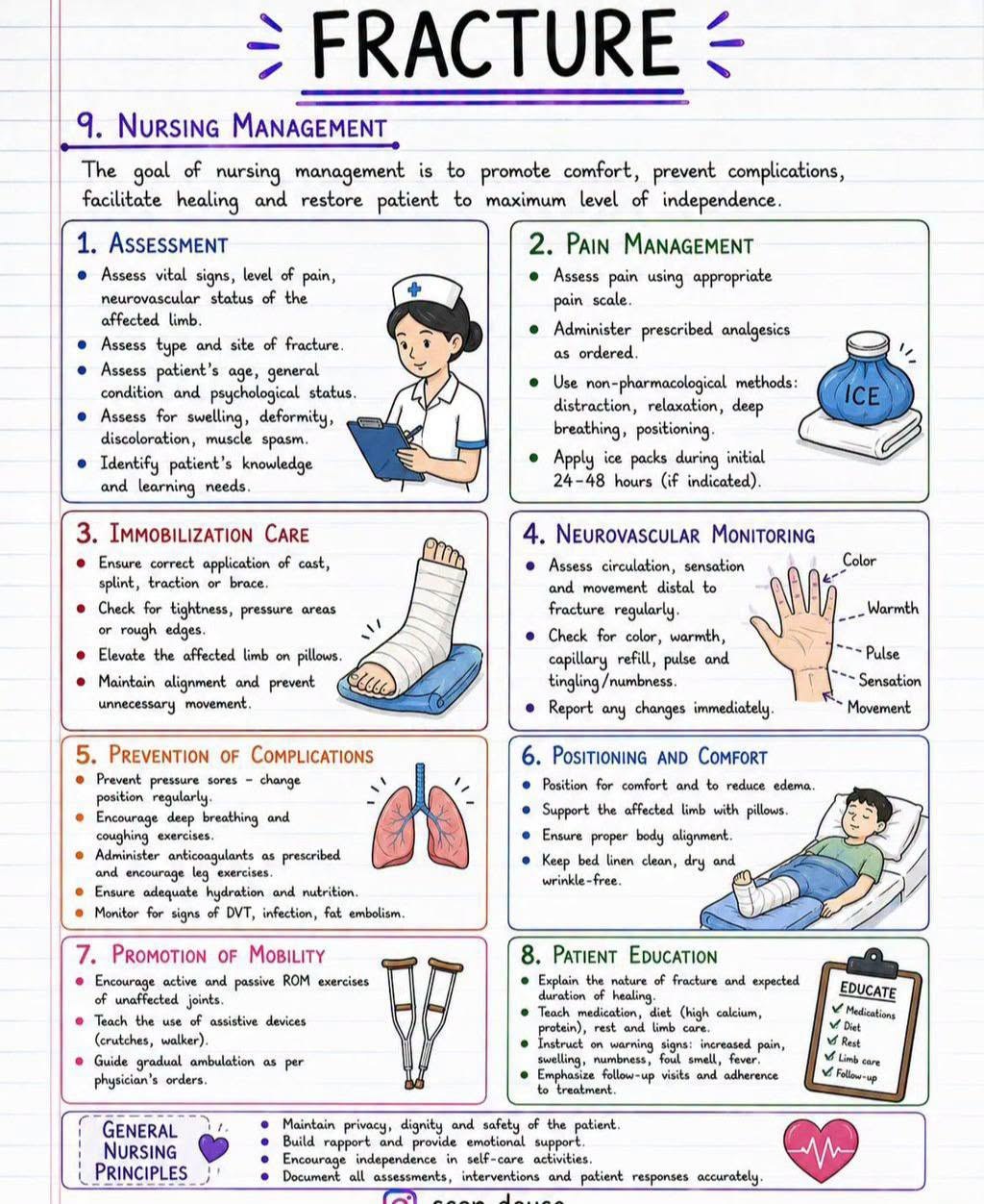
Part 7 — the eight nursing priorities, with neurovascular monitoring spelled out: color, warmth, pulse, sensation, movement. — swipe it sideways if it is cut off, or tap to open it full size.
Saved study graphics from your own collection. Each one is someone else’s work — check anything clinical against your course materials before you rely on it.