The Skin 🧬
Structure & function — the foundation page for the whole Skin & Wound set
Skin is not wrapping paper — it is the body's largest organ, about 1.5–2 m² of surface and roughly 15% of body weight. Every other page in this set (pressure injuries, wound care, frostbite, dermatitis, psoriasis, skin cancer) is a story about which layer got hurt and how deep it went. Learn this cross-section once and the rest becomes depth, not memorization.
📄 Simple Nursing original — opens in Drive →
THE LAYERS
STEP 1 · STRUCTUREOne drawing to rule the whole set — memorize this cross-section and every later page is just "how deep did the injury go?"
🔬 Master cross-section — full-thickness skin
🧬 Epidermis — the barrier, and it has no blood supply
Avascular. It is fed by diffusion from the dermis below. That is why a shallow scrape oozes clear fluid instead of bleeding, and why the epidermis alone can regenerate perfectly with no scar.
- Keratinocytes — 90%+ of the cells. Born at the bottom, pushed up, flatten, die, flake off. Full turnover ~28 days (faster in psoriasis — see NG-225).
- Melanocytes — make melanin, which shields DNA from UV. Everyone has about the same number; skin tone reflects how much pigment they make.
- Langerhans cells — the immune sentinels. They present antigen; they are the reason allergic contact dermatitis exists (NG-182).
- Merkel cells — light touch / discriminative sensation, sitting on the basal layer.
🔬 Zoom: the strata, and the 28-day escalator
🧬 Dermis — the workshop where everything lives
This is the layer with the blood, nerves, glands and follicles. Injuries that reach it hurt, bleed and can scar.
- Papillary dermis (thin, upper) — loose tissue + capillary loops that feed the epidermis; contains Meissner corpuscles.
- Reticular dermis (thick, lower) — dense collagen (tensile strength) and elastin (recoil = turgor). Stretch it too fast and you get striae.
- Holds the appendages: hair follicles, sebaceous glands, sweat glands — the cells that let partial-thickness wounds re-epithelialise from the middle outward.
- Two vascular plexuses: a deep cutaneous plexus and a superficial subpapillary plexus — the body's heat radiator.
🧬 Hypodermis — fat, insulation, and the drug depot
- Adipose + loose connective tissue. Insulation, shock absorption, energy store, anchors skin to fascia.
- Thickness varies enormously by site and body habitus — this is exactly why subcutaneous vs intramuscular injection depth is chosen by site and needle length.
- Thin fat over a bony prominence = the classic pressure injury set-up (NG-044).
- Poor perfusion here is why the cachectic, the very old and the very cold injure so fast.
⭐ The appendages — three glands people constantly mix up
| Structure | Empties into | Secretes | Where | Clinical hook |
|---|---|---|---|---|
| Eccrine sweat gland | Its own pore on the skin surface | Watery, salty sweat | Almost everywhere; densest palms, soles, forehead | The real cooling system — evaporation. Lost fluid + Na⁺ + K⁺ |
| Apocrine sweat gland | The hair follicle | Thicker, protein-rich fluid | Axillae, groin, areolae | Odorless until skin bacteria digest it → body odor; activates at puberty |
| Sebaceous gland | The hair follicle | Oily sebum | Everywhere except palms & soles; densest face, scalp, back | Blocked + inflamed = acne. Keeps the surface supple and slightly acidic |
Acid mantle: sebum + sweat keep the surface pH around 4.5–5.5. That acidity plus normal flora is real antimicrobial defense — which is why harsh soap, repeated scrubbing and incontinence-associated moisture all raise infection risk.
WHAT SKIN DOES
STEP 2 · FUNCTIONSix jobs. When you can name them, you can predict exactly what goes wrong when skin is lost.
🛡️ The six functions, drawn
🌡️ Thermoregulation — the two settings
Evaporation of sweat is what actually removes heat — sweat sitting on the skin does nothing until it evaporates. Humid air, occlusive dressings and heavy blankets all block it.
💥 What is lost when skin is lost
Every complication of a big wound or burn is just a function on this page failing:
- Barrier gone → infection, then sepsis.
- Water control gone → evaporative fluid loss, hypovolemia, hypothermia.
- Protein leaves in exudate → low albumin → more edema → slower healing.
- Heat control gone → the patient gets cold fast, especially in the OR or in an open wound.
- Sensation gone → repeat injury goes unnoticed (diabetic foot, neuropathy).
⚠️ Who has fragile skin — flag these on admission
LESION LANGUAGE
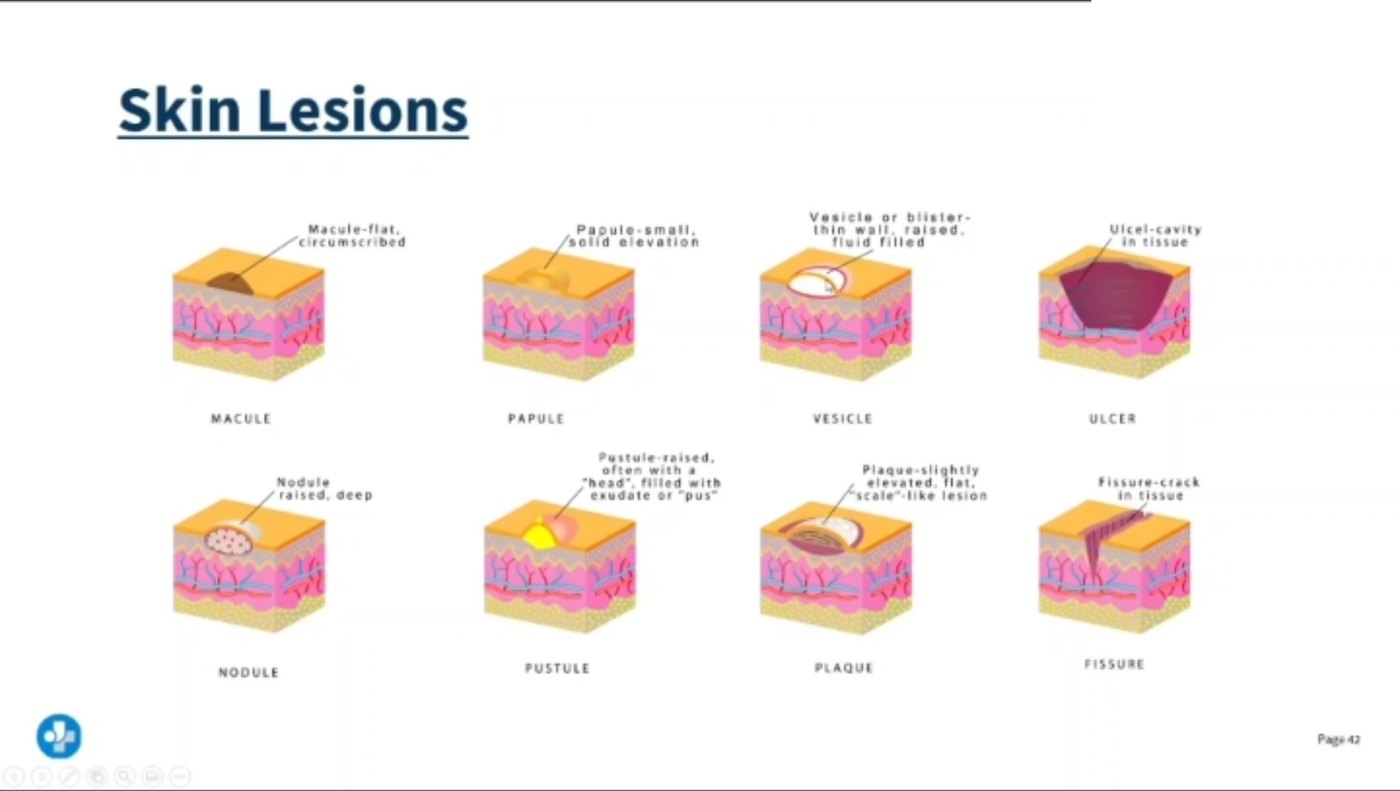
STEP 3 · DESCRIBE IT"Rash" is not an assessment. These nine words are the vocabulary every derm question is written in.
🔎 PRIMARY lesions — the first thing the skin does
🔎 SECONDARY lesions — what happens next (scratching, healing, breaking down)
Also learn: lichenification = thickened, leathery skin with exaggerated skin lines from chronic rubbing (the hallmark of long-standing atopic dermatitis — NG-182); atrophy = thin, translucent, "cigarette paper" skin (chronic topical steroids); scar = fibrous replacement tissue; keloid = scar that grows past the original wound edges.
🚨 The blanching test — the one that separates benign from serious
Press a red area with your finger, then release.
- Blanches (turns white, then re-reddens) = blood still inside vessels. Reactive hyperemia, mild erythema, sunburn.
- Does NOT blanch = blood has leaked outside the vessels, or tissue is already damaged. Non-blanchable erythema over a bony prominence is a Stage 1 pressure injury (NG-044).
- Non-blanching pinpoint red-purple spots = petechiae; larger flat patches = purpura; a big bruise = ecchymosis. Petechiae + fever is an escalate-now finding.
📝 How to describe a lesion so it counts as charting
Use the same six slots every time and you can chart any skin finding:
- Type — macule, papule, vesicle, ulcer…
- Size — measure in cm (length × width × depth), never "the size of a quarter"
- Color — and whether it blanches
- Location — anatomical landmark, and left/right
- Configuration — linear, annular (ring), grouped/clustered, target/iris, serpiginous
- Distribution — localized vs generalized, flexural (eczema) vs extensor (psoriasis), dermatomal (shingles), sun-exposed
ASSESS & PROTECT
STEP 4 · TECHNIQUEInspection, palpation, and the traps — especially in darker skin and in older adults.
🩺 Three bedside tests, drawn
⭐ Assessing color change in darker skin — high-yield and often taught badly
Erythema, cyanosis and jaundice can be genuinely hard to see in deeply pigmented skin. Do not rely on color alone — palpate and compare.
- Compare to the surrounding skin and the same site on the other side.
- Inflammation may look purple, violet, darker brown or ashen rather than pink or red.
- Palpate for the other three signs: warmth or coolness, induration (firmness), boggy/spongy texture, edema, and pain reported by the patient.
- Use natural or bright non-fluorescent light; check sclera, conjunctiva, oral mucosa, palms/soles and nail beds for pallor, cyanosis and jaundice.
- Stage 1 pressure injuries are under-detected in darker skin — this is a known equity gap, so lower your threshold and document what you palpate, not only what you see.
✅ The head-to-toe skin check — order and content
- Timing: a full skin assessment on admission (within the first 24 hours per most facility policy), then per policy and every shift for at-risk patients.
- Look — color, symmetry, lesions, hygiene, hair distribution, nails (clubbing, spooning, splinter hemorrhages).
- Touch — temperature (back of your hand), moisture, texture, turgor, edema (press over a bony area, grade pitting).
- Don't skip the hidden places: behind the ears, under breasts and skin folds, between toes, perineum, sacrum, heels, occiput, and under every medical device (tubing, oximeter probe, O₂ tubing, cast edge, cervical collar).
- Turn the patient — you cannot assess a sacrum you never looked at.
👵 Ageing skin vs young skin — why the same injury is worse at 85
- Flattened dermo-epidermal junction → the layers slide apart with shear; skin tears from tape and transfers.
- Less sebum and fewer sweat glands → dry, itchy skin (xerosis) and poorer heat tolerance.
- Fewer vessels + slower cell turnover → wounds heal more slowly and infect more easily.
- Less fat → bony prominences are barely padded.
- Turgor is unreliable as a hydration measure in older adults — use the sternum or forehead, and confirm with mucous membranes, intake/output and daily weights.
📏 Depth language that every later page uses
- Superficial / epidermal — epidermis only. Heals from the basal layer, no scar. (Sunburn, erosion, Stage 1.)
- Partial thickness — epidermis + part of the dermis. Painful (nerves exposed), moist, heals by re-epithelialising from the surviving hair follicles and glands. (Blister, Stage 2, superficial frostbite.)
- Full thickness — through the dermis into fat or deeper. No appendages left, so it must fill in with granulation tissue from the base and edges → scar, contracture, long healing. (Stage 3–4, deep frostbite, deep burns.)
Follow the depth story: NG-044 Pressure Injuries stages exactly this cross-section; NG-080 Wound Care turns depth into a dressing choice; NG-116 Frostbite is the cold version of the same depths.
💊 One derm drug that shows up constantly — isotretinoin
A vitamin A derivative used for severe, nodulocystic acne that failed other therapy.
- Pregnancy is the absolute priority. It is a potent teratogen. Prescribers and patients enroll in the manufacturer's risk-management program (iPLEDGE in the US): a negative pregnancy test before starting and before refills, and two forms of contraception used together.
- Report mood changes or suicidal thoughts immediately — psychiatric monitoring is part of the teaching.
- No extra vitamin A supplements (additive toxicity).
- Avoid tetracyclines concurrently — the combination is linked to raised intracranial pressure (report severe headache, visual changes, nausea/vomiting).
- Expect dry skin, dry eyes and dry lips: lubricating drops, may not tolerate contact lenses, and strict sun protection — the skin burns easily.
- Monitor lipids and liver enzymes; avoid alcohol.
✅ Universal skin protection — good for every patient on every page
- Keep it clean and dry — gentle pH-balanced cleanser, pat don't rub, moisturise while skin is still damp.
- Manage moisture — treat incontinence promptly, use a barrier cream/film; moisture-associated damage is not a pressure injury but sets one up.
- Never massage a reddened bony prominence — it damages already-injured microcirculation.
- Reposition and offload, float heels, use lift sheets, minimize head-of-bed elevation to limit shear.
- Feed the skin — adequate protein and calories, and fluids unless restricted. Track albumin/prealbumin (typical adult albumin reference band ≈ 3.5–5.0 g/dL; ranges vary by lab) as a nutrition/inflammation marker.
- Sun protection — broad-spectrum SPF 30+, reapply every 2 h (NG-204).
🗺️ How this page connects to the rest of the set
| Page | What it adds on top of this cross-section |
|---|---|
| NG-044 — Pressure Injuries | Redraws this exact cross-section with progressively deeper loss: Stage 1 → 4, plus unstageable and deep tissue injury; risk scoring and prevention |
| NG-080 — Wound Care | How wounds close (primary/secondary/tertiary intention), the healing phases, dressing choice, drains, dehiscence and evisceration |
| NG-116 — Frostbite | The same depths, caused by ice crystals and vasoconstriction; rewarming rules |
| NG-182 — Dermatitis | Uses the lesion vocabulary above: vesicles, weeping, lichenification, flexural vs contact-shaped distribution |
| NG-225 — Psoriasis | What happens when the 28-day keratinocyte escalator speeds up to a few days: silvery scaly plaques on extensor surfaces |
| NG-204 — Skin Cancer | What goes wrong in the basal layer (basal cell), the keratinocytes (squamous cell) and the melanocytes (melanoma) |