The complications — and every single one starts with back-pressure
NG-109GI · HEPATICADHD-friendly visual edition
What this page covers, and what it doesn't.NG-029 (Cirrhosis I) covered the pathophysiology, the causes and the
liver's four jobs. This page is the complications page: portal hypertension, ascites, esophageal varices
and the bleeding emergency, hepatic encephalopathy, coagulopathy, jaundice and SBP — what you SEE and what the
LABS show. NG-148 (Cirrhosis III) is the care page: procedures, drugs, diet
and teaching. Read this one first; almost everything on it grows out of a single sentence —
blood cannot get through the scarred liver, so it backs up.
🩸 Everything backs upBlood can't cross the scar → portal hypertension → ascites, varices, big spleen.
🎈 Ascites = 3 causesHigh portal pressure + low albumin + aldosterone the liver can't clear.
🚨 Vomiting blood?AIRWAY first — side-lying, suction ready. Then vitals and volume.
🧠 Ammonia ↑Confusion + asterixis = hepatic encephalopathy. Compare with the last shift.
🩸
PORTAL HYPERTENSION — THE ROOT OF EVERYTHING
STEP 1 · CAUSE
One blocked organ, four escape routes, and every escape route is a complication.
🪨 What the scar actually does to the blood flow
Cirrhosis replaces soft, spongy liver tissue with fibrous scar and regenerative
nodules. Scar tissue cannot be squeezed through, so the pressure in the portal vein climbs.
🧠 “The liver turns to rock.” Nothing flows through rock — so the river floods backwards.
🗺️ Where the blocked blood escapes to
🧠 “Water finds a way.” Esophagus, umbilicus, rectum, spleen — the four escape routes,
the four classic signs.
🧪 What portal hypertension IS
The portal vein normally runs at a low pressure. In cirrhosis the pressure rises; a portal pressure
gradient above roughly 10 mmHg is the level at which the classic complications start
to appear, and higher pressures carry a higher bleeding risk.
Blood that cannot get through the liver is forced into small collateral veins that were never built
to carry that volume — so they stretch, thin out, and eventually tear.
🧠 “Dam the river, flood the side streets.” Collaterals are the flooded side streets.
👀 What you can see at the bedside
Distended, tense abdomen with a fluid wave and shifting dullness (ascites).
Caput medusae — dilated veins radiating from the navel.
Splenomegaly — you may see it on imaging and the platelet count reflects it.
Hemorrhoids that are new or worsening.
Weight gain and increasing abdominal girth without eating more.
🧠 Measure girth at the SAME marked spot every day, or the number means nothing.
🩸 Splenomegaly → low platelets
Blood dams back into the spleen and it enlarges. An enlarged spleen sequesters and destroys
platelets, so the platelet count falls.
Now stack that on top of a liver that cannot make clotting factors: fewer platelets AND fewer clotting
factors at the same time. That is why a cirrhotic patient bruises from a blood-pressure cuff.
Position: semi-Fowler's or high Fowler's to let the diaphragm drop.
Never lie a patient with tense ascites flat for comfort — it makes the breathing worse.
🧠 “Big belly, sit them up.” Positioning is a real intervention here, not a comfort measure.
🦠 Spontaneous bacterial peritonitis (SBP)
Ascitic fluid is a warm pool of protein sitting in the abdomen. It can become infected without any
obvious source — no perforation, no surgery, nothing.
Fever, sometimes low-grade or absent in a very sick liver patient.
Abdominal pain or tenderness, rebound tenderness.
New or worsening confusion — often the first thing the nurse notices.
Worsening ascites that suddenly stops responding to treatment.
Diagnosis is by sampling the ascitic fluid; treatment is antibiotics per culture and protocol.
🧠 New confusion + fever + a big belly = think SBP. Report it, do not wait for the morning round.
✅ Nursing priorities for the ascitic abdomen
Position semi- to high Fowler's for breathing.
Daily weight, girth and strict I&O.
Low-sodium diet, and a fluid restriction if one is ordered.
Skin care — stretched, edematous abdominal skin breaks down and weeps.
Support the abdomen when the patient moves; teach them to splint when coughing.
Monitor for SBP — temperature, abdominal tenderness, mental status.
🧠 Sit up · weigh · measure · watch the skin · watch the temperature.
🚨
ESOPHAGEAL VARICES — THE EMERGENCY
STEP 3 · THE KILLER
This is the complication that kills people quickly, and the one every exam asks about.
💣 What varices are, and why they rupture
🧠 “A ticking time bomb of blood.” Thin-walled veins with the pressure of a whole
blocked circulation behind them, sitting on top of the airway.
🚨 FIRST ACTION when the patient vomits blood
A
AIRWAY. Turn the patient side-lying so blood drains out of the mouth instead of into the lungs. Suction at the bedside, oxygen on.
▼
B
BREATHING. Oxygen, monitor saturation, watch for aspiration.
▼
C
CIRCULATION. Vital signs, large-bore IV access, blood work and type and cross, prepare for volume and blood products.
▼
D
Call for help and prepare for urgent endoscopy. Keep the patient NPO.
Some questions ask “what is the FIRST action?” and the safest answer is the one that
protects the airway; others ask “what would the nurse do first” in a stable-sounding scenario, where
obtaining vital signs is the assessment answer. Read whether the patient is actively bleeding into
their mouth right now.
🧠 Blood in the mouth → turn them on their side. You cannot resuscitate a blocked airway.
❌ The two orders you QUESTION
A new nasogastric tube in a patient with known or suspected varices — a tube
passed blindly down the esophagus can tear a varix open. Question the order and speak to the provider.
Anything that causes straining — enemas that require bearing down, hard
suppositories, and any instruction to push.
Also question aspirin and NSAIDs unless specifically ordered, and any invasive oral or esophageal device.
🧠 “No tube down the pipe.” If the exam gives you an NG tube order in a varices patient,
that is the answer.
🚽 Why straining is so dangerous
Bearing down (the Valsalva maneuver) raises intra-abdominal and intrathoracic pressure. That pressure
is transmitted straight to the already-stretched varices.
Teach: do not strain with bowel movements, do not hold your breath when moving up the bed.
Stool softeners as ordered so that stools stay soft.
Also avoid heavy lifting and forceful coughing where possible.
🧠 “Don't push.” The exam wording is usually “avoid straining when having a bowel movement.”
👀 Recognizing a bleed before it is obvious
SIGN
WHAT IT MEANS
Hematemesis — bright red vomit
Active, brisk upper GI bleeding
Coffee-ground emesis
Blood that has been sitting in the stomach
Melaena — black tarry stool
Digested blood from an upper GI source
Rising pulse, falling BP
Losing volume — often before any visible blood
New confusion
Poor perfusion, and a protein load feeding ammonia production
🧠 Black tarry stool is old blood; bright red vomit is new blood. Both are emergencies here.
✅ Preventing the first bleed
Non-selective beta blockers as prescribed, to lower portal pressure. Check heart rate and blood
pressure against the hold parameters before every dose.
Endoscopic banding of known varices, done electively.
No alcohol, no NSAIDs, no straining, no blind NG tubes.
Teach the patient and family what hematemesis and melaena look like, and to call emergency services.
🧠 Prevention beats resuscitation. The beta blocker is doing quiet work every day.
🧠
HEPATIC ENCEPHALOPATHY
STEP 4 · THE BRAIN
Cloudy brain from ammonia. The nurse usually spots it before the lab does.
🧪 Where the ammonia comes from and where it ends up
🧠 Protein → ammonia → liver → urea → urine. Break the liver step and the ammonia goes to
the brain instead.
🖐️ Asterixis — how to test for it
🧠 “The liver flap.” Arms out, hands back, watch. It looks like a bird flapping its wings.
👀 The three key assessments
1
Ask the orientation questions. Name, date of birth, date, place. Ask them the same way every shift.
2
Extend the arms and watch the hands. Asterixis is the classic motor sign.
3
Compare with the previous shift's documentation and with recent ammonia levels — the direction of travel matters more than one number.
That trio is the answer to the classic select-all-that-apply question about detecting
hepatic encephalopathy.
🧠 “Ask · Extend · Compare.” Three assessments, one diagnosis.
📉 How it progresses
1
Subtle. Disturbed sleep pattern (awake at night, sleepy by day), mild forgetfulness, slight personality change. Families notice this first.
▼
2
Obvious. Lethargy, disorientation, asterixis, inappropriate or bizarre behavior.
▼
3
Marked. Very drowsy but rousable, confused, incoherent speech, cannot follow commands.
▼
4
Coma. Unresponsive. Airway and aspiration risk.
🧠 Sleep pattern first, coma last. Day–night reversal is a real early sign, not just “bad sleep”.
🔥 What sets it off
GI bleeding — a huge protein load dumped into the gut. This is the big one.
Constipation — ammonia sits in the bowel longer and is reabsorbed.
Infection, including SBP.
Dehydration and electrolyte disturbance, often from over-diuresis.
Sedatives and opioids, which the failing liver cannot clear.
A large protein meal in a patient who is already borderline.
Never give a sedative to “settle” a confused cirrhotic patient without a provider's assessment.
🧠 Bleed · block · bug · dry · drug. Five triggers to check whenever the mental status drops.
🛡️ Safety while they are confused
Fall precautions — bed low, call bell in reach, frequent rounding.
Aspiration precautions — assess swallowing before anything by mouth; keep them sitting up.
Reorient calmly and keep the environment consistent; involve family who know their baseline.
Skin and pressure care — a lethargic patient does not reposition themselves.
Document the mental status in the same words each time so the next nurse can compare.
🧠 Confused liver patient = fall risk + aspiration risk. Both, every time.
🧪
THE LIVER-FAILURE LAB PANEL
STEP 5 · LABS
Which ones go up, which go down, and — more usefully — why.
📊 Every value, and the reason behind it
🧠 “The liver's ABC goes DOWN: Albumin, Bile handling, Coagulation.
Everything the liver was supposed to REMOVE goes UP.”
🩸 Coagulopathy — two problems stacked
The liver makes most clotting factors and needs vitamin K to activate several of them. It also sits
upstream of a spleen that is now destroying platelets.
PT · PTT · INRAll PROLONGED — it takes longer to clot.
PlateletsLOW — trapped and destroyed in a big spleen.
Result: bruising from a blood-pressure cuff, bleeding gums, prolonged oozing from
any puncture, and a GI bleed that will not stop on its own.
Typical adult albumin is 3.5–5.0 g/dL. In cirrhosis it falls, and that single value
explains several findings at once:
Ascites and peripheral edema — nothing left to hold water in the vessels.
Low total calcium — much of the calcium in blood is carried bound to albumin, so the total falls
with it (typical calcium 9.0–10.5 mg/dL).
Drugs behave unpredictably — many are albumin-bound, so more free drug circulates.
🧠 Low albumin = low calcium reading + water in the wrong place.
🦴 Low calcium — the two classic signs
TROUSSEAU'S SIGNInflate a blood-pressure cuff above systolic for a few minutes → the hand and wrist go into carpal spasm.
CHVOSTEK'S SIGNTap over the facial nerve just in front of the ear → the facial muscles twitch.
Also watch for numbness and tingling around the mouth and in the fingers, muscle
cramps, and hyperactive reflexes.
🧠 “TrousseauS = Spasm of the hand. ChvosteK = Cheek.”
🧪 Enzymes vs function — a distinction that gets tested
TELLS YOU
TESTS
Liver cells are being DAMAGED
ALT, AST (leaked out of injured cells)
Liver FUNCTION is failing
Albumin, PT/INR, bilirubin, ammonia
Enzymes can even be near normal in end-stage cirrhosis — there are too few working liver cells left to
leak anything. A normal ALT does not mean a healthy liver.
🧠 Enzymes = damage. Albumin, INR and bilirubin = function. Function is what matters.
🎓 Select all that apply — the classic version
Which values are expected to be ELEVATED in worsening liver failure?
✅ Ammonia — the liver cannot convert it to urea.
✅ Bilirubin — it cannot be cleared into the bile.
✅ Prothrombin time (PT) / INR — fewer clotting factors, so it takes longer.
❌ Albumin — goes DOWN; the liver cannot make it.
❌ Calcium — goes DOWN, largely because albumin does.
🧠 The trick in this question is that albumin and calcium travel together, and they travel
in the OPPOSITE direction to everything else.
💛
JAUNDICE, ITCHING AND THE REST OF THE BODY
STEP 6 · HEAD TO TOE
The visible signs that make an exam question recognizable in one sentence.
💛 The jaundice cascade
🧠 “The Bile Bus has stopped running.” The pigment cannot get to the toilet, so it
goes into the skin instead.
🧍 Head-to-toe in one card
SYSTEM
WHAT YOU FIND
👁️ Eyes / skin
Yellow sclera and skin, spider angiomas, palmar erythema, easy bruising, scratch marks
🧠 Neuro
Confusion, day–night reversal, asterixis, lethargy → coma
🫁 Respiratory
Shortness of breath from a raised diaphragm; rapid shallow breathing
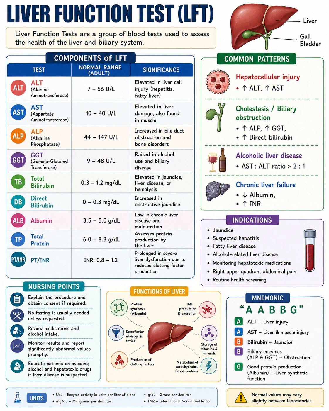
Every LFT with its adult reference range, and the pattern-matching that matters: hepatocellular versus cholestatic versus alcoholic versus chronic failure. — swipe it sideways if it is cut off, or tap to open it full size.
Saved study graphics from your own collection. Each one is someone else’s work — check anything clinical against your course materials before you rely on it.